| Listing 1 - 10 of 11 | << page >> |
Sort by
|
Book
ISBN: 3540783598 9783540783596 Year: 2008 Publisher: Heidelberg : Springer Medizin Verlag,
Abstract | Keywords | Export | Availability | Bookmark
 Loading...
Choose an application
- Reference Manager
- EndNote
- RefWorks (Direct export to RefWorks)
Eye Diseases --- Eye --- Fluorescein Angiography --- Fluorescence angiography --- diagnosis --- Diseases --- Diagnosis
Book
ISBN: 9781582557991 Year: 2010 Publisher: Baltimore (Md.) : Lippincott William & Wilkins,
Abstract | Keywords | Export | Availability | Bookmark
 Loading...
Choose an application
- Reference Manager
- EndNote
- RefWorks (Direct export to RefWorks)
Fluorescence angiography. --- Fluorescence. --- Fundus Oculi. --- Fundus oculi. --- Lipofuscin --- Lipofuscins. --- Ophthalmoscopy --- Retinal Diseases --- Physiology. --- Methods. --- Diagnosis.
Book
ISBN: 3540352244 Year: 2008 Publisher: Heidelberg : Springer Medizin,
Abstract | Keywords | Export | Availability | Bookmark
 Loading...
Choose an application
- Reference Manager
- EndNote
- RefWorks (Direct export to RefWorks)
Bei der technischen Weiterentwicklung von Angiographiesystemen konnte in den letzten Jahren ein enormer Fortschritt erzielt werden. Hierdurch hat sich die Bildqualität bei der Fluoreszein- und Indozyaningrün-Angiographie erheblich verbessert. Auch durch die wesentlich genaueren Möglichkeiten der Autofluoreszenz-Bestimmung können ganz neue Einblicke in die Pathogenese insbesondere von Makula- und Netzhauterkrankungen gewonnen werden. In dem neuen Fluoreszenzangiographie-Atlas von Dithmar und Holz werden die Grundlagen der Fluoreszein- und Indozyaningrün-Angiographie sowie der Autofluoreszenz-Bestimmung dargestellt. Die verschiedenen angiographischen Merkmale klinischer Krankheitsbilder werden anhand praxisrelevanter Fallbeispiele herausgearbeitet. Dabei wurde bei der Bildauswahl besonderes Augenmerk auf die Qualität sowie auf klar erkennbare, typische Veränderungen gelegt. Der Atlas bietet so einen Überblick über die facettenreichen angiographischen Befunde retinologischer Krankheitsentitäten und Differenzialdiagnosen. Auch der nicht selbst angiographierende Augenarzt wird von diesem Atlas in seinem pathophysiologischen und klinischen Verständnis profitieren.
Ophthalmology. --- Medicine. --- Medicine/Public Health, general. --- Eye --- Fluorescence angiography. --- Indocyanine green --- Diseases --- Diagnosis. --- Radiography. --- Diagnostic use.

ISBN: 1556427875 9781556427879 Year: 2008 Publisher: Thorofare (N.J.) : SLACK,
Abstract | Keywords | Export | Availability | Bookmark
 Loading...
Choose an application
- Reference Manager
- EndNote
- RefWorks (Direct export to RefWorks)
'Fundus Fluorescein and Indocyanine Green Angiography: A Textbook and Atlas' is a complete and detailed reference that comprehensively covers fluorescein angiography, the more recent and advancing indocyanine green angiography and their effectiveness in identifying and evaluating various retinal diseases. Prof. Amar Agarwal has teamed up with more than 30 retina specialists to provide eyecare professionals with the most current and essential information for the successful employment and accurate interpretation of these vital diagnostic tests. Sections inside the book include: Basics and principles Macular diseases Vascular retinal diseases Non vascular retinal diseases Optic nerve disorders This timely reference uses a unique textbook and atlas format to convey its key points and includes over 550 images and photographs to provide a visual representation of the disease process. Also included is an accompanying CD-ROM with over 35 minutes of supplemental video. Topics on video CD-ROM include: Fundus fluorescein angiography Indocyanine green angiography Optical coherence tomography Mirror telescopic IOL for macular degenerations With fluorescein and indocyanine green angiography continuing to grow as important and critical diagnostic tests, 'Fundus Fluorescein and Indocyanine Green Angiography: A Textbook and Atlas' will keep ophthalmologists, retina specialists, and optometrists on the cutting edge in today's fast-paced and ever-changing industry. Some topics covered inside include: Evolution of angiography Instrumentation Normal angiograms Macular holes Tumors OCT and spectral OCT
Coloring Agents --- Dyes in medical diagnosis. --- Fluorescein Angiography. --- Fluorescence angiography. --- Indocyanine Green --- Indocyanine green --- Retina --- Retinal Diseases --- Diagnostic use. --- Diseases --- Diagnosis.

ISBN: 1280610565 9786610610563 3540300619 3540300600 3642440851 Year: 2006 Publisher: New York, NY : Springer,
Abstract | Keywords | Export | Availability | Bookmark
 Loading...
Choose an application
- Reference Manager
- EndNote
- RefWorks (Direct export to RefWorks)
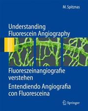
Fluorescein angiography is an indispensable tool in modern ophthalmology. It is founded on the evaluation of phenomena related to the behavior intravenously administered sodium fluorescein in the tissues of the ocular fundus. Commonly, texts on fluorescein angiography merely describe the typical pattern, quality, and quantity of the phenomena specific to a variety of vascular and other diseases of the retina and choroid. In contrast, the present book deals with the morphological prerequisites and structural changes on which the individual phenomena are based. This knowledge provides the retina specialist, the general ophthalmologist, and the basic scientist alike with valuable information toward understanding the nature and mechanism of the underlying disorders. The book represents a unique mixture of textbook and atlas, always combining concise statements in English, German, and Spanish on one page with didactically conceived explanatory illustrations on the facing page.
Fluorescence angiography. --- Ophthalmic photography. --- Eye --- Photography, Ophthalmic --- Photography in ophthalmology --- Medical photography --- Fluorescein angiography --- Fundus fluorescence photography --- Angiography --- Ophthalmic photography --- Photography --- Examination --- Blood-vessels --- Radiography --- Ophthalmology. --- Radiology, Medical. --- Science, Humanities and Social Sciences, multidisciplinary. --- Imaging / Radiology. --- Clinical radiology --- Radiology, Medical --- Radiology (Medicine) --- Medical physics --- Medicine --- Diseases --- Radiology. --- Radiological physics --- Physics --- Radiation
Book
ISBN: 1281070289 9786611070281 3540719946 3540719938 3642091199 Year: 2007 Publisher: Berlin : Springer,
Abstract | Keywords | Export | Availability | Bookmark
 Loading...
Choose an application
- Reference Manager
- EndNote
- RefWorks (Direct export to RefWorks)
During recent years, FAF (Fundus autofluorescence) imaging has been shown to be useful in various retinal diseases with regard to diagnostics, documentation of changes, identification of disease progression, and monitoring of novel therapies. Hereby, FAF imaging gives additional information above and beyond conventional imaging tools. This unique atlas provides a comprehensive and up-to-date overview of FAF imaging in retinal diseases. It also compares FAF findings with other imaging techniques such as fundus photograph, fluorescein- and ICG angiography as well as optical coherence tomography. General ophthalmologists as well as retina specialists will find this a very useful guide which illustrates typical FAF characteristics of various retinal diseases.
Retina --- Fluorescence angiography --- Retinal degeneration --- Diseases --- Imaging --- Diagnosis --- Posterior segment (Eye) --- Fluorescein angiography --- Fundus fluorescence photography --- Angiography --- Eye --- Ophthalmic photography --- Dystrophy, Retinal --- Macular degeneration --- Retinal dystrophy --- Degeneration (Pathology) --- Blood-vessels --- Radiography --- Degeneration --- Ophthalmology. --- Radiology, Medical. --- Imaging / Radiology. --- Clinical radiology --- Radiology, Medical --- Radiology (Medicine) --- Medical physics --- Medicine --- Radiology. --- Radiological physics --- Physics --- Radiation
Book
ISBN: 1281772925 9786611772925 3540794018 Year: 2008 Publisher: Heidelberg : Springer,
Abstract | Keywords | Export | Availability | Bookmark
 Loading...
Choose an application
- Reference Manager
- EndNote
- RefWorks (Direct export to RefWorks)
The technology of angiographic systems has been improved tremendously just within the past few years. This has allowed greatly increased levels of image resolution for both fluorescein and indocyanine green angiography. This new atlas by Dithmar and Holz covers the basic principles of the new methods for fluorescein- and indocyanine green-angiography, as well as the high resolution imaging of fundus autofluorescence. The angiographic signs of retinal and choroidal diseases are illustrated with images taken from a series of clinically relevant case examples that specifically illustrate the advantages of higher image resolution for the study of common retinochoroidal disorders. In so doing, this atlas offers an all-encompassing survey of the many angiographic signs in these disorders and their differential diagnoses. Clinicians in all subspecialties of ophthalmology can profit from a better understanding of these pathophysiological phenomena.
Eye --- Fluorescence angiography --- Diseases --- Diagnosis --- Fluorescein angiography --- Fundus fluorescence photography --- Angiography --- Ophthalmic photography --- Eyeball --- Eyes --- Visual system --- Face --- Photoreceptors --- Vision --- Blood-vessels --- Radiography --- Ophthalmology. --- Medicine. --- Medicine/Public Health, general. --- Clinical sciences --- Medical profession --- Human biology --- Life sciences --- Medical sciences --- Pathology --- Physicians --- Medicine --- Health Workforce
Book
Year: 2022 Publisher: Basel MDPI - Multidisciplinary Digital Publishing Institute
Abstract | Keywords | Export | Availability | Bookmark
 Loading...
Choose an application
- Reference Manager
- EndNote
- RefWorks (Direct export to RefWorks)
The purpose of this SI is to provide an overview of recent advances made in the methods used for tissue imaging and characterization, which benefit from using a large range of optical wavelengths. Guerouah et al. has contributed a profound study of the responses of the adult human brain to breath-holding challenges based on hyperspectral near-infrared spectroscopy (hNIRS). Lange et al. contributed a timely and comprehensive review of the features and biomedical and clinical applications of supercontinuum laser sources. Blaney et al. reported the development of a calibration-free hNIRS system that can measure the absolute and broadband absorption and scattering spectra of turbid media. Slooter et al. studied the utility of measuring multiple tissue parameters simultaneously using four optical techniques operating at different wavelengths of light—optical coherence tomography (1300 nm), sidestream darkfield microscopy (530 nm), laser speckle contrast imaging (785 nm), and fluorescence angiography (~800 nm)—in the gastric conduit during esophagectomy. Caredda et al. showed the feasibility of accurately quantifying the oxy- and deoxy-hemoglobin and cytochrome-c-oxidase responses to neuronal activation and obtaining spatial maps of these responses using a setup consisting of a white light source and a hyperspectral or standard RGB camera. It is interest for the developers and potential users of clinical brain and tissue optical monitors, and for researchers studying brain physiology and functional brain activity.
Public health & preventive medicine --- hemodynamic brain mapping --- metabolic brain mapping --- Monte Carlo simulations --- intraoperative imaging --- optical imaging --- hyperspectral imaging --- RGB imaging --- fluorescence imaging --- fluorescence angiography --- indocyanine green (ICG) --- optical coherence tomography (OCT) --- laser speckle contrast imaging (LSCI) --- esophagectomy --- gastric conduit --- Sidestream Darkfield Microscopy (SDF) --- multispectral --- broadband diffuse reflectance spectroscopy --- frequency-domain near-infrared spectroscopy --- dual-slope --- absorption spectra --- supercontinuum laser --- NIRS --- tissue optics --- diffuse optics --- near-infrared spectroscopy --- brain --- BOLD signal --- breath-holding --- cytochrome C oxidase --- n/a
Book
Year: 2022 Publisher: Basel MDPI - Multidisciplinary Digital Publishing Institute
Abstract | Keywords | Export | Availability | Bookmark
 Loading...
Choose an application
- Reference Manager
- EndNote
- RefWorks (Direct export to RefWorks)
The purpose of this SI is to provide an overview of recent advances made in the methods used for tissue imaging and characterization, which benefit from using a large range of optical wavelengths. Guerouah et al. has contributed a profound study of the responses of the adult human brain to breath-holding challenges based on hyperspectral near-infrared spectroscopy (hNIRS). Lange et al. contributed a timely and comprehensive review of the features and biomedical and clinical applications of supercontinuum laser sources. Blaney et al. reported the development of a calibration-free hNIRS system that can measure the absolute and broadband absorption and scattering spectra of turbid media. Slooter et al. studied the utility of measuring multiple tissue parameters simultaneously using four optical techniques operating at different wavelengths of light—optical coherence tomography (1300 nm), sidestream darkfield microscopy (530 nm), laser speckle contrast imaging (785 nm), and fluorescence angiography (~800 nm)—in the gastric conduit during esophagectomy. Caredda et al. showed the feasibility of accurately quantifying the oxy- and deoxy-hemoglobin and cytochrome-c-oxidase responses to neuronal activation and obtaining spatial maps of these responses using a setup consisting of a white light source and a hyperspectral or standard RGB camera. It is interest for the developers and potential users of clinical brain and tissue optical monitors, and for researchers studying brain physiology and functional brain activity.
hemodynamic brain mapping --- metabolic brain mapping --- Monte Carlo simulations --- intraoperative imaging --- optical imaging --- hyperspectral imaging --- RGB imaging --- fluorescence imaging --- fluorescence angiography --- indocyanine green (ICG) --- optical coherence tomography (OCT) --- laser speckle contrast imaging (LSCI) --- esophagectomy --- gastric conduit --- Sidestream Darkfield Microscopy (SDF) --- multispectral --- broadband diffuse reflectance spectroscopy --- frequency-domain near-infrared spectroscopy --- dual-slope --- absorption spectra --- supercontinuum laser --- NIRS --- tissue optics --- diffuse optics --- near-infrared spectroscopy --- brain --- BOLD signal --- breath-holding --- cytochrome C oxidase --- n/a
Book
Year: 2022 Publisher: Basel MDPI - Multidisciplinary Digital Publishing Institute
Abstract | Keywords | Export | Availability | Bookmark
 Loading...
Choose an application
- Reference Manager
- EndNote
- RefWorks (Direct export to RefWorks)
The purpose of this SI is to provide an overview of recent advances made in the methods used for tissue imaging and characterization, which benefit from using a large range of optical wavelengths. Guerouah et al. has contributed a profound study of the responses of the adult human brain to breath-holding challenges based on hyperspectral near-infrared spectroscopy (hNIRS). Lange et al. contributed a timely and comprehensive review of the features and biomedical and clinical applications of supercontinuum laser sources. Blaney et al. reported the development of a calibration-free hNIRS system that can measure the absolute and broadband absorption and scattering spectra of turbid media. Slooter et al. studied the utility of measuring multiple tissue parameters simultaneously using four optical techniques operating at different wavelengths of light—optical coherence tomography (1300 nm), sidestream darkfield microscopy (530 nm), laser speckle contrast imaging (785 nm), and fluorescence angiography (~800 nm)—in the gastric conduit during esophagectomy. Caredda et al. showed the feasibility of accurately quantifying the oxy- and deoxy-hemoglobin and cytochrome-c-oxidase responses to neuronal activation and obtaining spatial maps of these responses using a setup consisting of a white light source and a hyperspectral or standard RGB camera. It is interest for the developers and potential users of clinical brain and tissue optical monitors, and for researchers studying brain physiology and functional brain activity.
Public health & preventive medicine --- hemodynamic brain mapping --- metabolic brain mapping --- Monte Carlo simulations --- intraoperative imaging --- optical imaging --- hyperspectral imaging --- RGB imaging --- fluorescence imaging --- fluorescence angiography --- indocyanine green (ICG) --- optical coherence tomography (OCT) --- laser speckle contrast imaging (LSCI) --- esophagectomy --- gastric conduit --- Sidestream Darkfield Microscopy (SDF) --- multispectral --- broadband diffuse reflectance spectroscopy --- frequency-domain near-infrared spectroscopy --- dual-slope --- absorption spectra --- supercontinuum laser --- NIRS --- tissue optics --- diffuse optics --- near-infrared spectroscopy --- brain --- BOLD signal --- breath-holding --- cytochrome C oxidase
| Listing 1 - 10 of 11 | << page >> |
Sort by
|

 Search
 Feedback
 About UniCat
  Help
Help News
News