
 Search
 Feedback
 About UniCat
  Help
Help News
News
| Listing 1 - 9 of 9 |
Sort by
|
Book
ISBN: 9782294779909 Year: 2022 Publisher: Issy-les-Moulineaux : Elsevier Masson,
Abstract | Keywords | Export | Availability | Bookmark
 Loading...
Choose an application
- Reference Manager
- EndNote
- RefWorks (Direct export to RefWorks)
La TEP-TDM est désormais une technique d'imagerie essentielle en oncologie. Permettant d'étudier l'activité métabolique des tissus grâce à un traceur radioactif elle a transformé en profondeur les modalités de prise en charge des patients présentant un cancer. Mais les possibilités qu'elle offre lui permettent également de jouer un rôle important dans l'élaboration de diagnostics de pathologies non cancéreuses. L'ouvrage rappelle d'abord l'anatomie des structures présentes au niveau de l'ensemble du corps (squelette peau muscles graisse et ganglions). Puis classées par région anatomique les pathologies peuvent être comparées grâce à plus de 800 clichés de haute qualité. La pathologie maligne les tumeurs bénignes la pathologie infectieuse la pathologie dégénérative et éventuellement les maladies de système sont ainsi largement documentées afin de souligner ce qui peut aider au diagnostic différentiel. Sont ainsi successivement abordés : le cerveau le cou et la face le thorax les seins l'abdomen l'appareil urinaire les surrénales le pelvis féminin la prostate les testicules et la pédiatrie. Cette organisation recoupe la pratique quotidienne lors de l'interprétation des examens TEP-TDM. Cet ouvrage destiné principalement à un public de médecins nucléaires de radiologues d'oncologues et d'hématologues confirmé ou en devenir constitue le premier ouvrage de référence en langue française sur le sujet.
Tomography, Emission. --- Tomography. --- Neoplasms --- Tomographie par émission. --- Scanographie. --- Cancer --- Tomographie par émission de positons couplée à la tomodensitométrie. --- diagnostic imaging. --- diagnosis. --- Diagnostic. --- Imagerie.
Book
ISBN: 9781461400905 1461400902 Year: 2011 Publisher: New York ; London : Springer,
Abstract | Keywords | Export | Availability | Bookmark
Loading...Choose an application
- Reference Manager
- EndNote
- RefWorks (Direct export to RefWorks)
Tomography, Emission. --- Tomography. --- Cancer --- Positron-Emission Tomography and Computed Tomography --- Neoplasms --- Tomographie par émission --- Scanographie --- radionuclide imaging. --- Tomographie
Book
ISBN: 9780781788977 0781788978 Year: 2010 Publisher: Philadelphia : Wolters Kluwer Health/Lippincott William & Wilkins,
Abstract | Keywords | Export | Availability | Bookmark
Loading...Choose an application
- Reference Manager
- EndNote
- RefWorks (Direct export to RefWorks)
Brain Diseases --- Tomography, Emission-Computed --- Brain --- Tomography, Emission. --- Cerveau --- Tomographie par émission --- diagnosis --- Atlases. --- methods --- Tomography. --- Tomographie --- Tomographie par émission
Book
ISBN: 1416061126 9781416061120 Year: 2008 Publisher: Philadelphia : W. B. Saunders Company,
Abstract | Keywords | Export | Availability | Bookmark
Loading...Choose an application
- Reference Manager
- EndNote
- RefWorks (Direct export to RefWorks)
Book

ISBN: 9791030300383 Year: 2016 Publisher: Montpellier : Sauramps Medical,
Abstract | Keywords | Export | Availability | Bookmark
Loading...Choose an application
- Reference Manager
- EndNote
- RefWorks (Direct export to RefWorks)
Divisé en 2 parties, ce livre discute l?apport de la tomoscintigraphie osseuse en pathologie du pied et de la cheville. La première partie est consacrée à la pathologie : les principales pathologies de la cheville et du pied visibles en scintigraphie osseuse sont expliquées sous forme synthétique, avec les données essentielles de l?apport de la scintigraphie. Les attentes précises des chirurgiens sont mises en valeur. De nombreuses scintigraphies osseuses d?intérêt clinique sont présentées. Certaines pathologies fréquentes ou utiles à connaître mais non diagnostiquées par la scintigraphie osseuse seront décrites également (névrome de Morton par exemple). La deuxième partie est consacrée à l?anatomie : les os, les articulations et toutes les structures anatomiques pertinentes et suffisantes à l?interprétation d?une scintigraphie osseuse de la cheville et du pied sont présentés au moyen de dessins légendés. A la fin des années 2000, l?arrivée des machines hybrides, couplant une gamma-caméra à une tomodensitométrie, a ouvert un vaste champ de perspectives pour les médecins nucléaires. Comme leurs confrères radiologues au début des années 1990, les médecins nucléaires découvrent des pathologies qui leur sont méconnues et des foyers de fixation jusqu?ici invisibles. Cet ouvrage, écrit par des médecins nucléaires et des chirurgiens orthopédiques spécialistes de la cheville et du pied, constitue une aide à l?apprentissage de cette nouvelle sémiologie. Points clés : - Présentation de la longue expérience en prescription de scintigraphie osseuse de deux chirurgiens orthopédiques spécialistes de la cheville et du pied. - Présentation et discussion autour de cas cliniques de haut intérêt clinique. - Abord à la fois pathologique et anatomique, essentiel pour la réalisation et l?interprétation d?une tomoscintigraphie osseuse.
Imagerie médicale. --- Imagerie pour le diagnostic. --- Tomographie par émission. --- Os --- Cheville (anatomie) --- Pied --- Tomoscintigraphie. --- Cheville. --- Pied. --- Ankle --- Foot --- Tomography, Emission --- Diagnostic imaging --- Cheville --- Tomographie par émission --- Imagerie pour le diagnostic --- Maladies --- Diagnostic. --- Scintigraphie. --- Pathology --- Diseases --- Radionuclide imaging --- Maladie --- Scintigraphie
Multi
ISBN: 9783319400709 9783319400686 Year: 2017 Publisher: Cham Springer International Publishing
Abstract | Keywords | Export | Availability | Bookmark
Loading...Choose an application
- Reference Manager
- EndNote
- RefWorks (Direct export to RefWorks)
This book offers a wide-ranging and up-to-date overview of the basic science underlying PET and its preclinical and clinical applications in modern medicine. In addition, it provides the reader with a sound understanding of the scientific principles and use of PET in routine practice and biomedical imaging research. The opening sections address the fundamental physics, radiation safety, CT scanning dosimetry, and dosimetry of PET radiotracers, chemistry and regulation of PET radiopharmaceuticals, with information on labeling strategies, tracer quality control, and regulation of radiopharmaceutical production in Europe and the United States. PET physics and instrumentation are then discussed, covering the basic principles of PET and PET scanning systems, hybrid PET/CT and PET/MR imaging, system calibration, acceptance testing, and quality control. Subsequent sections focus on image reconstruction, processing, and quantitation in PET and hybrid PET and on imaging artifacts and correction techniques, with particular attention to partial volume correction and motion artifacts. The book closes by examining clinical applications of PET and hybrid PET and their physiological and/or molecular basis in conjunction with technical foundations in the disciplines of oncology, cardiology and neurology, PET in pediatric malignancy and its role in radiotherapy treatment planning. Basic Science of PET Imaging will meet the needs of nuclear medicine practitioners, other radiology specialists, and trainees in these fields.
Physical methods for diagnosis --- klinische chemie --- PET (positron-emission tomography) --- PET/CT --- pneumologie --- radiologie --- medische beeldvorming --- Nuclear medicine. --- Radiology. --- Tomography, Emission. --- Radiologie. --- Médecine nucléaire. --- Tomographie par émission.

ISBN: 0824708849 Year: 2003 Volume: 19 Publisher: New York : M. Dekker,
Abstract | Keywords | Export | Availability | Bookmark
Loading...Choose an application
- Reference Manager
- EndNote
- RefWorks (Direct export to RefWorks)
Affective disorders --- Brain --- Tomography, Emission. --- Troubles affectifs --- Cerveau --- Tomographie par émission --- Mood Disorders --- Magnetic Resonance Imaging. --- Magnetic Resonance Spectroscopy. --- Tomography, Emission-Computed. --- Imaging. --- Magnetic resonance imaging. --- Diagnosis. --- Imagerie --- Imagerie par résonance magnétique --- Diagnostics --- anatomy & histology. --- diagnosis.
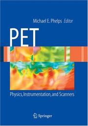
ISBN: 1280724269 9786610724260 0387349464 0387323023 1441921834 Year: 2006 Publisher: New York : Springer,
Abstract | Keywords | Export | Availability | Bookmark
Loading...Choose an application
- Reference Manager
- EndNote
- RefWorks (Direct export to RefWorks)
Derived from the critically acclaimed reference PET : Molecular Imaging and Its Biological Applications, edited by Michael E. Phelps, Ph.D., this handbook provides a clear and concise introduction to the physics and instrumentation aspects of PET imaging. Comprised of select material from the parent volume, this book is gives the reader a solid understanding of PET fundamentals, including how PET data are collected and formed into an image. Other topics include basic physics, detector technology used in modern PET scanners, data acquisition, 3-D reconstruction, and methods for evaluating the performance of PET systems. A variety of modern PET imaging systems is discussed, ranging from those designed for clinical services and research to those used in small-animal imaging. As the importance of PET imaging in nuclear medicine and radiology grows, the need for a reliable resource increases. PET: Physics, Instrumentation, and Scanners is a handy guide to this important field.
Tomography, Emission. --- Diagnosis --- Data processing. --- Computerized emission tomography --- Emission tomography --- PET (Tomography) --- PET-CT (Tomography) --- Positron emission tomography --- Positron emission transaxial tomography --- Radionuclide tomography --- Scintigraphy, Tomographic --- Tomography, Radionuclide --- Diagnostic imaging --- Positrons --- Radioisotope scanning --- Data processing --- Emission --- Tomographie par émission --- Nuclear medicine. --- Radiology, Medical. --- Nuclear Medicine. --- Imaging / Radiology. --- Clinical radiology --- Radiology, Medical --- Radiology (Medicine) --- Medical physics --- Atomic medicine --- Radioisotopes in medicine --- Medical radiology --- Radioactive tracers --- Radioactivity --- Physiological effect --- Tomographie par émission. --- pet skanning --- røntgendiagnostik --- postion-emission tomografi --- røntgenundersøgelser --- radiografi --- nuklearmedicin --- Radiology. --- Radiological physics --- Physics --- Radiation --- Tomographie par émission.
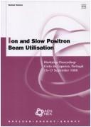
ISBN: 1280034076 9786610034079 9264172998 9264170251 Year: 1999 Publisher: Paris : OECD Publishing,
Abstract | Keywords | Export | Availability | Bookmark
Loading...Choose an application
- Reference Manager
- EndNote
- RefWorks (Direct export to RefWorks)
The use of ion beams in nuclear research is well established, with many facilities and networks of experts active in the field. Applications for ion beams are expanding, in particular in the development of new materials, biotechnology and the creation of new isotopes. Positron beams are likewise a very powerful tool for observing and influencing microscopic material structures, as well as for medical diagnosis. The combined utilisation of ion and positron beams is expected to open up new horizons in the areas of material science and biotechnology. These proceedings provide an overview of the latest developments in this field, and highlight areas for future international co-operation.
Nuclear Energy --- Ion bombardment --- Positron beams --- Radioactive nuclear beams --- Tomography, Emission --- Materials science --- Physics --- Physical Sciences & Mathematics --- Electricity & Magnetism --- Industrial applications --- Portugal --- Computerized emission tomography --- Emission tomography --- PET (Tomography) --- PET-CT (Tomography) --- Positron emission tomography --- Positron emission transaxial tomography --- Radionuclide tomography --- Scintigraphy, Tomographic --- Tomography, Radionuclide --- Beams, Radioactive nuclear --- Nuclear beams, Radioactive --- Radioactive beams --- Beams, Positron --- Beams, Ion --- Bombardment, Ion --- Impact, Ion --- Ion beams --- Ion impact --- Ionic bombardment --- Diagnosis --- Diagnostic imaging --- Positrons --- Radioisotope scanning --- Particle beams --- Collisions (Nuclear physics) --- Ions --- Data processing --- Emission --- Bombardement ionique --- Faisceaux de positons --- Faisceaux radioactifs --- Tomographie par émission --- Science des matériaux --- Congresses. --- Congresses --- Applications industrielles --- Congrès
| Listing 1 - 9 of 9 |
Sort by
|