| Listing 1 - 10 of 32 | << page >> |
Sort by
|
Book
Year: 1963 Publisher: Springfield, Ill. : Thomas,
Abstract | Keywords | Export | Availability | Bookmark
 Loading...
Choose an application
- Reference Manager
- EndNote
- RefWorks (Direct export to RefWorks)
Book
Year: 2017 Publisher: Bruxelles: UCL. Faculté de pharmacie et des sciences biomédicales,
Abstract | Keywords | Export | Availability | Bookmark
 Loading...
Choose an application
- Reference Manager
- EndNote
- RefWorks (Direct export to RefWorks)
The purpose of this brief is twofold. On the one hand, it highlights the place that Thyrogen occupies in the treatment of well differentiated thyroid cancer by distinguishing its two indications. It allows to situate it in the management of the cancer but also to highlight its non-inferiority as well as the advantages, in particular in terms of cost-effectiveness and quality of life, compared to another existing method which is thyroid hormone withdrawal. On the other hand, this work also deals with the production of Thyrogen and recombinant proteins in general in CHO mammalian cells. L'objectif de ce mémoire est double. D'une part, il met en évidence la place qu'occupe le Thyrogen® dans le traitement du cancer de la thyroïde bien différencié en distinguant ses deux indications. Il permet de le situer dans la prise en charge du cancer mais également de mettre en lumière sa non-infériorité ainsi que ses avantages, notamment en termes de coût-efficacité et qualité de vie, par rapport à une autre méthode existante qui est l'arrêt du traitement frénateur par la lévothyroxine. D'autre part, ce travail traite également de la production du Thyrogen® et des protéines recombinantes en général dans des cellules mammifères de type CHO.
Book
Year: 2000 Publisher: Bruxelles: UCL,
Abstract | Keywords | Export | Availability | Bookmark
 Loading...
Choose an application
- Reference Manager
- EndNote
- RefWorks (Direct export to RefWorks)
The thyrotrophin receptor (TSHr) plays a critical role in the physiopathology of the thyroid. As self-antigen capable to be recognized by self-antibodies, he’s involved into the development of autoimmune thyroid diseases. Its molecular cloning has allowed to develop experimental models on animals. The genetic immunization of outbred NMRI mice with the cDNA construct encoding the human TSHr takes part in these studies and tempts to produce a satisfying experimental model of the Graves’ disease. After the immunization, all mice responded by producing antibodies capable of recognizing the TSHr. Five out of twenty-nine female mice showed biological evidence of hyperthyroidism with elevated total T4 and suppressed TSH levels. The serum of these mice contained thyroid stimulating activity. In contrast, only one male out of thirty had moderately elevated serum total T4 with undetectable TSH values. The hyperthyroid animals had goiters witextensive lymphocytic infiltration characteristic of a TH2 immune response. In addition, they displayed ocular sings reminiscent of Graves’ ophtalmopathy, including edema, deposit of amorphous material and cellular infiltration of their extra-ocular muscles. These results demonstrate that genetic immunization of outbred NMRI mice with the human TSHr provides the most convincing animal model of Graves disease available to date.
On the other hand, the TSHr associated with thyroglobulin (Tg), iodinated Tg and thyroid peroxydase (TPO) are four differentiation markers of the thyroid cells and their immunohistochemistry detection can be useful for differential diagnosis. We have studied the evolution of their expression in different thyroid tumors : hot adenoma, cold adenoma, follicular carcinoma, papillary carcinoma and anaplastic carcinoma. Iodinated Tg is the first marker to totally disappear in the carcinoma, his detection is therefore useful to establish a differential diagnosis of the malignant tumors with regard to benign tumors. The analysis of TPO expression allows to discriminate the malignant tumors, seeing that the TPO disappears in the follicular carcinomas and persists in the papillary carcinoma. Its detection is therefore very useful because of these types of carcinoma have two very different prognosis. Tg disappears in the anaplastic carcinomas where TSHr is still expressed. Therefore, the immunohistochemical detection of TSHr could allow to determine the origin of very indiferrentiated thyroid tumors or metastasis. Although our study is still preliminary and that its diagnosis value should be supported by an increase if the case numbers and by a quantitative analysis, it could provide complementary informations useful for the clinicians Le récepteur à la TSH (rTSH) joue un rôle central dans la physiopathologie de la thyroïde. En tant qu’autoantigène susceptible d’être reconnu par des autoanticorps, il est impliqué dans l’étiopathogénie des maladies auto-immunitaires thyroïdiennes. Son clonage moléculaire a permis de réaliser de nombreuses études en vue de l’élaboration de modèles de thyroïdites chez l’animal. L’immunisation génétique des souris non consanguines NMRI avec le cDNA codant pour le rTSH humain fait partie de ces études et tente de constituer un modèle expérimental satisfaisant de la thyroïdite de Graves-Basedow. Suite à l’immunisation, toutes les souris produisent des anticorps capables de reconnaître le rTSH de T4 élevés et des taux de TSH effondrés. L’analyse du sérum de ces souris a démontré clairement une activité stimulante de la thyroïde. Par contre, seule une souris immunisée mâle sur trente a un taux de T4 modérément élevé associé à un taux en TSH effondré. Les animaux hyperthyroïdiens présentent un goitre associé ç une infiltration lymphocytaire étendue, caractéristique d’une réponse immunitaire de type TH2. En outre, ces animaux présentent des signes oculaires rappelant l’ophtalmopathie décrite dans la maladie de Graves-Basedow, incluant de l’œdème, un dépôt de matériel amorphe et une infiltration cellulaire des muscles extra-oculaires. Ces résultats démontrent que l’immunisation génétique des souris NMRI avec le rTSH humain fournit actuellement le modèle animal le plus convaincant de la maladie de Graves-Basedow.
D’autre part, le rTSH tout comme la thyroglobuline (Tg), la Tg fortement iodée et la thyroperoxydase (TPO) sont quatre marqueurs de différentiation de la thyroïde, dont la détection immunohistochimique peut être utilisée à des fins diagnostiques. Nous avons étudié l’évolution de leur expression dans différentes pathologies tumorales de la thyroïde : les nodules chauds, les nodules froids, les carcinomes folliculaires, les carcinomes papillaires et les carcinomes anaplasiques. La Tg fortement iodée est le premier marqueur à disparaître totalement dans les carcinomes, sa détection s’avère donc utile pour le diagnostic différentiel des carcinomes différenciés par rapport aux adénomes. Pair ailleurs, la TPO permet de discriminer les carcinomes différenciés, puisqu’elle disparaît dans les carcinomes folliculaires, alors qu’elle persiste dans les carcinomes papillaires. Sa détection est donc très utile car ces deux carcinomes différenciés ont des pronostics très différents. La Tg disparaît dans les carcinomes anaplasiques om persiste l’expression du rTSH. La détection immunohistochimique du rTSH devrait donc permettre de déterminer la nature thyroïdienne de tumeurs très indifférenciées ou de métastases. Bien que cette étude soit encre préliminaire et que sa valeur diagnostique devra être étayée par une augmentation de nouveaux cas et appuyée par des méthodes quantitatives, elle apporte néanmoins de nouvelles indications qui assureront un complément d’information aux cliniciens

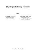
ISBN: 0890048010 Year: 1983 Publisher: New York Raven Press
Abstract | Keywords | Export | Availability | Bookmark
 Loading...
Choose an application
- Reference Manager
- EndNote
- RefWorks (Direct export to RefWorks)
Dissertation
Year: 1990 Publisher: 's-Gravenhage Pasmans
Abstract | Keywords | Export | Availability | Bookmark
 Loading...
Choose an application
- Reference Manager
- EndNote
- RefWorks (Direct export to RefWorks)
Thyrotropin --- Acromegaly --- Thyroid Gland --- Thyrotropin-Releasing Hormone --- blood --- physiopathology --- pharmacology
Dissertation
Year: 1982 Publisher: Groningen : Van Denderen,
Abstract | Keywords | Export | Availability | Bookmark
 Loading...
Choose an application
- Reference Manager
- EndNote
- RefWorks (Direct export to RefWorks)
Hyperthyroidism --- Prolactin --- Thyrotropin --- Physiopathology. --- Secretion.
Dissertation
Year: 1982 Publisher: Groningen Van Denderen
Abstract | Keywords | Export | Availability | Bookmark
 Loading...
Choose an application
- Reference Manager
- EndNote
- RefWorks (Direct export to RefWorks)
Thyrotropin --- Prolactin --- Hyperthyroidism --- secretion --- physiopathology
Dissertation
Abstract | Keywords | Export | Availability | Bookmark
 Loading...
Choose an application
- Reference Manager
- EndNote
- RefWorks (Direct export to RefWorks)
Thyrotropin --- Glycoproteins --- Physiopathology --- Glycoproteins. --- Physiopathology.
Book
ISBN: 0897664973 9780897664974 Year: 1989 Volume: 553 Publisher: New York, NY : New York Academy of Sciences,
Abstract | Keywords | Export | Availability | Bookmark
 Loading...
Choose an application
- Reference Manager
- EndNote
- RefWorks (Direct export to RefWorks)
Thyrotropin-Releasing Hormone --- Thyrotropin releasing factor --- Physiological effect --- Congresses --- Thyrotropin Releasing Hormone --- THYROTROPIN RELEASING HORMONE --- Congresses. --- congresses. --- Thyrotropin releasing hormone --- Thyrotropin-Releasing Hormone - congresses --- Thyrotropin releasing factor - Physiological effect - Congresses
Book
Abstract | Keywords | Export | Availability | Bookmark
 Loading...
Choose an application
- Reference Manager
- EndNote
- RefWorks (Direct export to RefWorks)
Thyroid Gland --- Thyroid Hormones --- Thyrotropin --- physiology
| Listing 1 - 10 of 32 | << page >> |
Sort by
|

 Search
 Feedback
 About UniCat
  Help
Help News
News