| Listing 1 - 10 of 22 | << page >> |
Sort by
|
Book
Year: 2016 Publisher: Bruxelles: UCL. Faculté de pharmacie et des sciences biomédicales,
Abstract | Keywords | Export | Availability | Bookmark
 Loading...
Choose an application
- Reference Manager
- EndNote
- RefWorks (Direct export to RefWorks)
Spermatogenesis is the formation process of spermatozoa from the spermatogonia. It takes place i n the testis, more particularly in the seminiferous tubules. The Sertoli cell is essential for the spermatogenesis because it is the physic and metabolic support of the germ cells. It provides protection and a number of nutrient s, growth factors and other substrates required for the survival and the differentiation. It receives endocrine and paracrine influences which make it possible to mature and differentiate during the puberty and consequently start up the spermatogenesis. The man fertility can decline due to gonadotoxic treatments, more specifically the chemotherapy and radiotherapy which can reduce the pool of germ stem cells. In the prepubertal boy, the spermatogenesis is not initiated yet and the only way to preserve fertility is the cryopreservation of a little fragment of testis tissue which contains spermatogonia. The ln vitro maturation could be a solution to obtain the differentiation of spermatogonial into spermatozoa. The aim of this master thesis is to study the different maturation and functional factors of the Sertoli cell in an organotypic culture of immature human testicular tissue in order to determine if this Sertoli cell can support the in vitro spermatogenesis processThe tissue fragments of 1mm3 of prepubertal patients are put in an organ culture during 139 days. Two culture media are used: one contains testosterone and the other contains the human choriogonadotropin (hCG, a LH-like) inducing the production of endogenous testosterone. Additionally, the two media are supplemented with follicle-stimulating hormone, mimicking the hormonal changes of the puberty. The tissue fragments are removed at specific time. They are embedded in paraffin and cut. They are colored with Hemalum-eosin in order to analyze the integrity of the seminiferous tubules (ST). The integrity is good (60% of the ST are considered as « good integrity »).lmmunochemistries (IHC) are used to bring out the factors of maturity or immaturity of the the Sertoli cell. The secretion of the anti-mullërian hormone (AMH) decreases during the culture, mimicking the peripubertal physiology of the maturation of the Sertoli cell. The proliferation (ki67) of the Sertoli cell increases, then decreases, which could also correspond to the maturation profile of the Sertoli cell. The Glial-derived neurotrophic factor, a functional factor of the Sertol i cell, is also is detected throughout the culture. Assays in the supernatants also allow characterizing the maturation and the functionality of the Sertoli cell. The AMH rate (by and enzymatic assay) decreases, confirming the results of the IHC. The measurement of the Dehydrogenase lactate (LDH, made by photometry) rate is rather high at the beginning of the culture and becomes almost zero at the 1Oth day, showing that there is little necrosis in the culture. Finally, a testosterone detection (by bioluminescence) in the medium supplemented with hCG show that the Leydig cells produce testosterone. ln our organotypic culture of immature testis, Sertoli cells show signs of maturation and functionality. l n the present culture system, any germ cell differentiation was observed. The absence of some growth factors with an in vivo systemic origin or functional disability of Sertoli cells in culture can be the cause and request further analysis. La spermatogenèse est le processus de formation des spermatozoïdes à partir de spermatogonies. Il se déroule au sein du tissu testiculaire, plus particulièrement dans les tubules séminifères. La cellule de Sertoli est une cellule capitale pour la spermatogenèse car elle sert de support physique et métabolique à la cellule germinale en lui fournissant une protection et un grand nombre de nutriments, facteurs de croissance et autres substances propices à sa survie et différenciation. Elle reçoit des influences endocrines et paracrines qui lui permettent de maturer et de se différencier lors de la puberté et ainsi d'enclencher le début de la spermatogenèse. La fertilité de l'homme peut se trouver diminuée par des traitements gonadotoxiques, plus particulièrement par les agents chimiothérapeutiques et radiothérapeutiques qui peuvent réduire la réserve de cellules germinales. Chez le garçon prépubère, pour qui la spermatogenèse n'est pas encore initiée, le seul moyen de préserver la fertilité avant ces traitements est la cryopréservation d'un fragment testiculaire contenant des spermatogonies. La maturation in vitro du tissu peut être une solution envisageable en vue d'obtenir la différenciation des spermatogonies en spermatozoïdes. Le but de ce mémoire est d'étudier différents facteurs de maturation et de fonctionnalité de la cellule de Sertoli dans une culture organotypique de tissu testiculaire immature humain afin de déterminer si cette dernière est capable de supporter le processus de la spermatogenèse in vitro. Des fragments de 1m m3 de tissu testiculaire de patient s prépubères sont mi s en culture organotypique durant 139 jours. Deux milieux de culture sont utilisés : l’un contenant de la testostérone et l’autre l’hormone chorionique gonadotrope humaine (hCG, une substance LH-like) induisant la production de testostérone endogène par action sur les celllules de Leydig. Les deux milieux sont, de surcroit, supplémentés en hormone folliculostimulante, mimant les changements hormonaux de la puberté. Les fragments, retirés à des jours précis, sont enrobés en bloc de paraffine et coupés. Ils sont colorés en hémalun-éosine afin d'analyser l’intégrité des tubules séminifères (TS). Celle-ci s'est révélée être bonne (60% des TS en moyenne sont considérés commeintègres).Des immunohistochimies (IHC) sont réalisées pour mettre en évidence des facteurs de maturité ou d'immaturité de la Sertoli. La sécrétion de l l'hormone antimüllérienne (AMH) diminue au cours de la culture, montrant une maturation de la cellule de Sertoli. La prolifération (Ki67) des cel lules de Sertoli augmente puis diminue, mimant la physiologie péripubère vers la maturation de la cellule de Sertoli. Un facteur de la fonctionnalité de la cellule de Sertoli , le Gl ial-derived neurotrophic factor, est détecté tout au long de la culture.Des dosages dans le surnageant permettent également de caractériser la maturation et la fonctionnalité de la cellule de Sertoli. Le taux d'AMH (par dosage enzymatique) diminue, confirmant les résultats de l 'IHC. La mesure du taux de lactate déshydrogénase (LDH, réalisé par photométrie) est assez haute au début de culture et devient quasi nul au 10ième jour de culture, montrant qu'il y a peu de nécrose dans la culture. Enfin, une détection de testostérone (par bioluminescence) dans le milieu supplémenté en hCG permet de montrer que les cellules de Leydig produisent de la testostérone.Dans notre culture organotypique de tissu testiculaire immature, les cellules de Sertoli montrent des signes de maturation et de fonctionnalité. Dans le système de culture actuel, aucune différenciation des cellules germinales n'est observée. L'absence de certains facteurs de croissance d’origine systémique in vivo ou une incapacité fonctionnelle des cellules de Sertoli en culture peuvent en être les causes et leur détermination nécessite donc un complément d’investigatio
Sertoli Cells --- Sertoli Cells --- Testicular Neoplasms --- Cryopreservation

ISBN: 9780126477511 0126477515 9780080492285 0080492282 1281005592 9781281005595 9786611005597 Year: 2005 Publisher: Amsterdam Boston Elsevier Academic Press
Abstract | Keywords | Export | Availability | Bookmark
 Loading...
Choose an application
- Reference Manager
- EndNote
- RefWorks (Direct export to RefWorks)
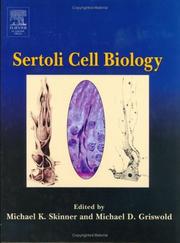
Sertoli cells assist in the production of sperm in the male reproductive system. This book provides a state-of-the-art update on the topic of sertoli cells and male reproduction. It addresses such highly topical areas as stem cells, genomics, and molecular genetics, as well as provides historical information on the discovery of this type of cell, and the pathophysiology of male infertility.* Presents the state-of-the-art research on topics such as stem cell research, transplantation and genomics* Includes contributions from leaders in the field, including several members of the Nat
Sertoli cells --- Spermatozoa. --- Physiology.
Dissertation
Year: 1988 Publisher: S.l. s.n.
Abstract | Keywords | Export | Availability | Bookmark
 Loading...
Choose an application
- Reference Manager
- EndNote
- RefWorks (Direct export to RefWorks)
Aging --- Inhibins --- Sertoli Cells --- physiology --- secretion
Book
ISBN: 012417048X 0124170471 9780124170483 9780124170476 Year: 2015 Publisher: Amsterdam, Netherlands
Abstract | Keywords | Export | Availability | Bookmark
 Loading...
Choose an application
- Reference Manager
- EndNote
- RefWorks (Direct export to RefWorks)
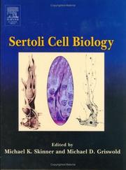
Sertoli Cell Biology, Second Edition summarizes the progress since the last edition and emphasizes the new information available on Sertoli/germ cell interactions. This information is especially timely since the progress in the past few years has been exceptional and it relates to control of sperm production in vivo and in vitro.
- Fully revised
- Written by experts in the field
- Summarizes 10 years of research
- Contains clear explanations and summaries
- Provides a summary of references over the last 10 years
Sertoli cells --- Physiology. --- Cells of Sertoli --- Sertoli's cells --- Sustentacular cells --- Cells --- Spermatozoa --- Testis
Dissertation
Year: 1987 Publisher: Utrecht Elinkwijk
Abstract | Keywords | Export | Availability | Bookmark
 Loading...
Choose an application
- Reference Manager
- EndNote
- RefWorks (Direct export to RefWorks)
Sertoli Cells --- Receptor, Insulin --- Follicle Stimulating Hormone --- Insulin --- drug effects --- metabolism --- pharmacology
Dissertation
Abstract | Keywords | Export | Availability | Bookmark
 Loading...
Choose an application
- Reference Manager
- EndNote
- RefWorks (Direct export to RefWorks)
Testis --- Testis --- Spermatogenesis --- Sertoli Cells --- drug effects --- growth & development --- physiology --- cytology

ISBN: 1281005592 9786611005597 0080492282 0126477515 9780126477511 9780080492285 9781281005595 Year: 2005 Publisher: Amsterdam Boston Elsevier Academic Press
Abstract | Keywords | Export | Availability | Bookmark
 Loading...
Choose an application
- Reference Manager
- EndNote
- RefWorks (Direct export to RefWorks)
Sertoli cells assist in the production of sperm in the male reproductive system. This book provides a state-of-the-art update on the topic of sertoli cells and male reproduction. It addresses such highly topical areas as stem cells, genomics, and molecular genetics, as well as provides historical information on the discovery of this type of cell, and the pathophysiology of male infertility.* Presents the state-of-the-art research on topics such as stem cell research, transplantation and genomics* Includes contributions from leaders in the field, including several members of the Nat
Sertoli cells --- Spermatozoa. --- Physiology. --- Male gametes --- Sperm --- Gametes --- Semen --- Cells of Sertoli --- Sertoli's cells --- Sustentacular cells --- Cells --- Spermatozoa --- Testis
Dissertation
Abstract | Keywords | Export | Availability | Bookmark
 Loading...
Choose an application
- Reference Manager
- EndNote
- RefWorks (Direct export to RefWorks)
Pituitary Gland --- Proteins --- Sertoli Cells --- Gonadotropin-Releasing Hormone --- Follicle Stimulating Hormone --- Inhibins --- drug effects --- isolation & purification --- metabolism --- analysis --- secretion --- physiology
Book
ISBN: 0702016195 9780702016196 Year: 1992 Volume: 6,2 Publisher: London : Baillière-Tindall,
Abstract | Keywords | Export | Availability | Bookmark
 Loading...
Choose an application
- Reference Manager
- EndNote
- RefWorks (Direct export to RefWorks)
Human histology. Human cytology --- Physiology: reproduction & development. Ages of life --- Testis --- Cytology --- Physiology --- Testis - Cytology --- Testis - Physiology --- TESTIS --- SERTOLI CELLS --- SPERMATOGENESIS --- INFERTILITY, MALE --- STEROIDS --- FSH --- INHIBIN --- PHYSIOLOGY
Dissertation
ISBN: 9789058676214 Year: 2007 Publisher: Leuven Universitaire Pers Leuven
Abstract | Keywords | Export | Availability | Bookmark
 Loading...
Choose an application
- Reference Manager
- EndNote
- RefWorks (Direct export to RefWorks)
Androgenen zijn essentieel voor spermatogenese. De mechanismen via dewelke ze hun effect op kiemcelontwikkeling uitoefenen blijven echter slecht begrepen. Alhoewel aan het begin van dit project de doelwitcel(len) die de effecten van androgenen op spermatogenese mediëren nog niet onomstotelijk geïdentificeerd was (waren), werden Sertoli cellen algemeen beschouwd als de voornaamste kandidaten gezien hun nauwe morfologische en functionele interacties met de zich ontwikkelende kiemcellen en het feit dat ze de androgeenreceptor op een stadiumafhankelijk manier tot expressie brengen tijdens de spermatogenese. De ontwikkeling - tijdens het verloop van deze studies - van een muismodel met een Sertoli celspecifieke uitschakeling van de androgeenreceptor (SCARKO), en de observatie dat in deze muizen de spermatogenese geblokkeerd is tijdens de meiose, toont ondubbelzinnig aan dat de androgeenreceptor in Sertoli cellen een centrale rol speelt in de regeling van de kiemcelontwikkeling. Om de mechanismen van androgeenwerking in Sertoli cellen te onderzoeken, hebben we eerst transfectiestudies met androgeen-responsieve promoter-reporter constructen uitgevoerd en hebben we microarraytechnologie aangewend om naar endogene androgeengeregelde genen te zoeken in geïsoleerde Sertoli cellen. Jammer genoeg was de voornaamste conclusie uit deze studies dat Sertoli cellen in belangrijke mate aan androgeengevoeligheid inboeten wanneer ze geïsoleerd en daarna in cultuur worden gebracht. Desalniettemin konden we enkele endogene genen identificeren die in beperkte mate antwoordden op androgenen. Bovendien antwoordden ook de promoter-reporter constructen, na cotransfectie met een exogeen androgeenreceptor plasmide, met een matige maar reproduceerbare verhoging in expressie op een behandeling met androgenen. Vermeldenswaardig is de bevinding dat de androgeenresponsen die onder deze condities werden waargenomen, maximaal waren bij androgeenconcentraties (~10-9 M) die duidelijk lager zijn dan deze vereist om spermatogenese te onderhouden. Na de succesvolle ontwikkeling van SCARKO muizen in ons laboratorium, werd dit unieke in vivo model gebruikt voor verdere studies omtrent de moleculaire mechanismen via dewelke androgenen spermatogenese beïnvloeden via Sertoli cellen. Uit een vergelijking van de genexpressie in testes van 10 dagen oude controle- en SCARKO-muizen door middel van microarraytechnologie, bleek dat 692 genen differentiëel tot expressie komen. Achtentwintig van deze genen vertoonden een meer dan tweemaal lagere expressie in SCARKO- vs. controle-testes (verder aangeduid als sterk neerwaarts geregeld) wat erop wijst dat deze genen sterk opwaarts geregeld zijn door androgenen. Van 3 van deze genen werd eerder aangetoond dat ze essentiëel zijn voor spermatogenese en voor 5 ervan bestonden er al directe of indirecte aanwijzingen voor androgeenresponsiviteit in de testis of in andere doelwitorganen. Twaalf genen waren sterk opwaarts geregeld in SCARKO-testes en bijgevolg vermoedelijk neerwaarts geregeld door androgeenactie in Sertoli cellen. Voor een selectie van 9 van de 28 genen die sterk neerwaarts geregeld waren in SCARKO-testes kon het verschil in genexpressie ook via kwantitatieve RT-PCR worden aangetoond en kon de androgeenregeling worden bevestigd in een onafhankelijk experimenteel model. Interessant was dat de rat homologen van 6 van deze geselecteerde genen ook androgeengeregeld bleken te zijn in rat testis en dat 4 van deze laatsten zelfs op androgenen antwoordden in rat Sertoli cellen in cultuur. Alles samengenomen ondersteunen deze resultaten de kracht van onze benaderingswijze om genen te identificeren die de effecten van androgenen op spermatogenese via Sertoli cellen mediëren. Daarenboven hebben functionele analyses en studies verder bouwend op de microarraydata serine protease inhibitoren en enzymes betrokken bij ganglioside biosynthese naar voren geschoven als belangrijke doelwitten voor androgeenactie in Sertoli cellen, wat erop wijst dat androgenen spermatogenese zouden kunnen beïnvloeden via effecten op tubulaire herstructureringen, celjunctie dynamica and cel-celcommunicatie. Samengevat kunnen we stellen dat onze genexpressiestudies op dag 10 de hypothese ondersteunen dat androgenen spermatogenese beïnvloeden via beperkte effecten op een waaier van verschillende genen eerder dan dat ze de expressie van één of meerder sleutelgenen zouden aan/uitschakelen. Deze effecten op genexpressie lijken echter toegespitst te zijn op een welbepaalde set van moleculaire processen zoals tubulaire herstructurering, celjunctie dynamica en cel-celcommunicatie. Verdere studies zijn aangewezen om het fysiologische effect van androgenen op elk van deze processen in meer detail te onderzoeken. Daarenboven zijn genexpressiestudies op jongere leeftijden verreist om na te gaan of de verschillen in genexpressie die werden waargenomen op dag 10 primaire en directe effecten van androgenen weergeven of dat ze een gevolg zijn van vroegere effecten van androgenen op de expressie van een andere en mogelijk beperktere set van genen. Androgens have clearly been shown to be essential for spermatogenesis. The mechanisms by which they exert their effects on germ cell development, however, have remained elusive. Although at the initiation of this project the target cell(s) which would mediate the effects of androgens on spermatogenesis was (were) not unambiguously identified, Sertoli cells were generally considered prime candidates given their intimate morphological and functional interactions with the developing germ cells and the fact that they express the AR in a stage-dependent manner during spermatogenesis. The development - during the course of these studies - of a mouse model with a Sertoli cell-selective knockout of the AR, and the demonstration that these mice (SCARKO) display a spermatogenic arrest in meiosis, unequivocally demonstrates that the AR in Sertoli cells plays a pivotal role in the control of germ cell development. To explore the mechanisms of androgen action in Sertoli cells, we first performed transfection studies with androgen-responsive promoter-reporter constructs and used microarray analysis to search for endogenous androgen-regulated genes in isolated Sertoli cells. Unfortunately, the major conclusion of these studies was that Sertoli cells loose many aspects of their androgen responsiveness upon isolation and culture. Nevertheless, we could identify a couple of endogenous genes with a limited response to androgens and, after cotransfection with an exogenous AR plasmid, also the promoter-reporter constructs responded to androgen treatment with a moderate but reproducible increase in expression. Interestingly, the androgen responses observed under these conditions were maximal at concentrations of androgens (~10-9 M) that are markedly lower than those required to maintain spermatogenesis. After the successful development of SCARKO mice in our laboratory, this unique in vivo model was used to further study the molecular mechanisms by which androgens affect spermatogenesis through Sertoli cells. By comparing gene expression in testes of 10-day-old control and SCARKO mice using microarray technology, 692 genes were found to be differentially expressed. Twenty eight of these genes displayed a more than twofold lower expression in SCARKO vs. control testes (further indicated as strongly downregulated) suggesting that these genes are strongly upregulated by androgens. Three of these genes were previously shown to be essential for spermatogenesis and for five of them direct or indirect evidence for androgen responsiveness existed in the testis or in other target tissues. Twelve genes were strongly upregulated in SCARKO testes and accordingly supposedly downregulated by androgen action in Sertoli cells. For a selection of 9 of the 28 genes which were strongly downregulated in SCARKO testes, differential expression was also shown by Q-RT-PCR and androgen regulation was confirmed in an independent experimental model. Interestingly, the rat homologues of 6 of these selected genes, also proved to be androgen regulated in the rat testis and 4 of the latter even responded to androgens in cultured rat Sertoli cells. Altogether these results support the power of our approach to identify genes mediating the effects of androgens on spermatogenesis through Sertoli cells. In addition, functional analyses and follow-up studies on the microarray data have put forward serine protease inhibitors and enzymes involved in ganglioside biosynthesis as major targets of androgen action in Sertoli cells, suggesting that androgens might influence spermatogenesis through effects on tubular restructuring events, cell junction dynamics and cell-cell communication. In conclusion, our gene expression studies on day 10 support the contention that androgens influence spermatogenesis through limited effects on the expression of an array of different genes in Sertoli cells rather than switching on/off the expression of one or several key genes. These diverse effects on gene expression, however, appear to be directed towards a confined set of molecular processes including tubular restructuring, cell junction dynamics and cell-cell communication. Future studies are warranted to examine the physiological effect of androgens on each of these processes in more detail. Moreover, gene expression studies at younger ages will be required to investigate if the differences in gene expression observed at day 10 represent primary and direct effects of androgens or if they are secondary to earlier effects of androgens on the expression of a different and possibly more limited set of genes. Androgenen (bv. testosteron) zijn mannelijke geslachtshormonen die geproduceerd worden in de teelbal. Naast het onderhouden van de mannelijke geslachtskenmerken, zijn zij cruciaal voor het onderhouden van de zaadcelproductie (spermatogenese) in de volwassen man. De teelbal is dus naast het belangrijkste productieorgaan óók het belangrijkste doelwitorgaan van androgenen. De mechanismen via dewelke androgenen de werking van cellen beïnvloeden zijn goed bestudeerd in accessoire geslachtsklieren zoals de prostaat. Testosteron bindt er aan een specifieke receptor, de androgeenreceptor. Deze geactiveerde receptoren gaan vervolgens in de celkern bepaalde genen stimuleren of juist afremmen. Merkwaardig genoeg beschikken de zich ontwikkelende zaadcellen (kiemcellen) niet over deze androgeenreceptor. Androgenen beïnvloeden spermatogenese dus vermoedelijk indirect, d.w.z. via andere cellen. Reeds bij het begin van dit project werd verondersteld dat Sertoli cellen, die de kiemcellen ondersteunen tijdens hun ontwikkeling, een belangrijk doelwit zijn voor androgenen in de controle van de spermatogenese. De ontwikkeling van een muismodel, tijdens het verloop van deze studies, waarin de androgeenreceptor specifiek is uitgeschakeld in Sertoli cellen (SCARKO muizen), heeft deze veronderstelling onomstotelijk bevestigd aangezien de spermatogenese in deze dieren wordt geblokkeerd tijdens de meiose. Over de mechanismen via dewelke androgenen hun effecten uitoefenen en over de genen die hierbij betrokken zijn, was echter nog weinig geweten. In deze thesis hebben wij daarom de mechanismen van androgeenwerking in Sertoli cellen onderzocht, eerst in geïsoleerde Sertoli cellen en na de ontwikkeling van de SCARKO muizen in dit unieke in vivo model. De studies in geïsoleerde Sertoli cellen leverden eerder teleurstellende resultaten op, maar toch konden we in dit in vitro model een aantal endogene
Sertoli cells --- Androgens --- Physiological effect --- Spermatogenesis --- Academic collection --- 618 --- 612.43 --- 612.43 Endocrine physiology. Internal secretions. Ductless glands. Hormones. Endocrinology --- Endocrine physiology. Internal secretions. Ductless glands. Hormones. Endocrinology --- 618 Gynaecology. Obstetrics --- Gynaecology. Obstetrics --- Theses
| Listing 1 - 10 of 22 | << page >> |
Sort by
|

 Search
 Feedback
 About UniCat
  Help
Help News
News