| Listing 1 - 10 of 27 | << page >> |
Sort by
|
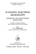
ISBN: 0123470501 Year: 1971 Publisher: London Academic Press
Abstract | Keywords | Export | Availability | Bookmark
 Loading...
Choose an application
- Reference Manager
- EndNote
- RefWorks (Direct export to RefWorks)
Biological microscopy --- Scanning electron microscope --- Congresses --- Scanning electron microscopy --- -Electron microscopy --- Congresses. --- scanning electron microscopy ( SEM ) --- -Congresses --- Scanning electron microscopes --- Electron microscopy

ISBN: 0521085314 9780521085311 Year: 1972 Publisher: Cambridge: Cambridge university press,
Abstract | Keywords | Export | Availability | Bookmark
 Loading...
Choose an application
- Reference Manager
- EndNote
- RefWorks (Direct export to RefWorks)
Optics. Quantum optics --- Scanning electron microscopes --- 537.533.35 --- Scanning electron microscope --- Electron microscopes --- Electron microscopy --- 537.533.35 Electron microscopy --- Monograph

ISBN: 3540061029 Year: 1973 Publisher: Berlin Springer
Abstract | Keywords | Export | Availability | Bookmark
 Loading...
Choose an application
- Reference Manager
- EndNote
- RefWorks (Direct export to RefWorks)
Dissertation
Year: 2019 Publisher: Liège Université de Liège (ULiège)
Abstract | Keywords | Export | Availability | Bookmark
 Loading...
Choose an application
- Reference Manager
- EndNote
- RefWorks (Direct export to RefWorks)
The imminent challenge in geometallurgical characterization of today’s ore deposits is its inherent heterogeneity and complexity which demands fast, resource-efficient, robust, and cost-effective means of measurement and analysis. To address this concern, mining operations all over the world are incorporating SEM-based automated mineralogy systems. Next generation platforms have been developed to bring out these technologies from the laboratories unto the field for on-site mineralogical characterization. Its widespread use have established its significance in the mining industry, however, cross-validation of results between technologies has not been well studied. This study aims to compare and validate results gathered from two SEM platforms: the ZEISS SIGMA 300 Gemini (FEG) with the ZEISS MinSCAN (W-filament) both are coupled with the ZEISS Mineralogic Mining system. Three sets of mixed Cu ore samples (feed, concentrate, and tails) from the Kansanshi deposit were analyzed exploring the effects of SEM operating conditions and sample preparation. The work focused on comparison of the modal mineralogy. The quality of results were assessed by its repeatability and cross-validated with chemical assay (AR-AAS). The limitations of these SEM-based systems were defined and solutions were proposed involving correlative microscopy. The feasibility of employing machine learning algorithms in image classification techniques were proposed to improve data acquisition process and accuracy of results of automated mineralogy systems. This study establishes the effect of the mineral recipe or SIP to the accuracy of results of a quantitative mineralogical analysis. Expertise in the deposit mineralogy as well as in the capabilities and limitations of the SEM is crucial in achieving reliable results. Cross-validation of back-calculated Mineralogic Cu and Fe grades from modal mineralogy with the measured AR-AAS chemical analysis shows that the error is minimized with the ZEISS SIGMA on samples prepared using carbon black. However, it must be noted that not all numerical values can be directly compared between the results and should be treated merely as indications of accuracy. Combined features from images obtained from the optical microscope (RGB) and the SEM (BSE) showed the least error in classification using a support vector machine algorithm. The segmented image shows mineral domains where EDX analysis can be performed on a set amount of points veering away from the pixel-by-pixel full mapping acquisition mode. This methodology can be developed and applied to automated mineralogy image processing systems as a domain-based acquisition mode. This has the potential to improve quantitative mineralogical analysis results accuracy while reducing acquisition time and resource costs.

ISBN: 1281139904 9786611139902 0387729720 0387729704 1489995846 Year: 2008 Publisher: New York : Springer,
Abstract | Keywords | Export | Availability | Bookmark
 Loading...
Choose an application
- Reference Manager
- EndNote
- RefWorks (Direct export to RefWorks)
Field-emission, low-voltage scanning electron microscopy (LVSEM) is a field that has grown tremendously in recent years because is offers the optimal method for viewing complex surfaces at high resolution and in three dimensions. However, even though the instrumentation required to get good results at low beam voltage has become increasingly available, there has been a lag in its application to biological specimens. What seemed to be missing was volume that combined both the theory and practice of using this equipment in an optimal manner with a thorough treatment of biological specimen preparation. Biological Low-Voltage Scanning Electron Microscopy is the first book to address both of these aspects of biological LVSEM. After providing a thorough description of the unique advantages and the operating constraints related to operating a scanning electron microscope at low beam voltage, the remainder of book focuses on the the best way to image all types of plant and animal cells and covers specimens that range from macromolecules to the surfaces revealed by de-embedding resin-embedded samples. Advanced specimen preparation techniques such as cryo-LVSEM, and immuno-gold-LVSEM are fully covered, as is x-ray microanalysis at low beam voltage and live-time stereo imaging. The preparative protocols provided represent the distilled essence of the experience of a group of world-renowned authors who have, for many decades, been instrumental in developing and applying new approaches to LVSEM to support their own biological research.
Low-voltage scanning electron microscopy. --- Field emission cathodes. --- Scanning electron microscopes. --- Scanning electron microscope --- Electron microscopes --- Cathodes --- Field emission --- Low voltage systems --- Scanning electron microscopy --- Microscopy. --- Zoology. --- Cytology. --- Microbiology. --- Biological Microscopy. --- Cell Biology. --- Microbial biology --- Biology --- Microorganisms --- Cell biology --- Cellular biology --- Cells --- Cytologists --- Natural history --- Animals --- Analysis, Microscopic --- Light microscopy --- Micrographic analysis --- Microscope and microscopy --- Microscopic analysis --- Optical microscopy --- Optics --- Cell biology.

ISBN: 0750302356 Year: 1993 Publisher: Bristol : Institute of Physics,
Abstract | Keywords | Export | Availability | Bookmark
 Loading...
Choose an application
- Reference Manager
- EndNote
- RefWorks (Direct export to RefWorks)
Alloys --- -Grain boundaries --- Metals --- -Scanning electron microscopes --- #KVIV:BB --- Scanning electron microscope --- Electron microscopes --- Metallic elements --- Chemical elements --- Ores --- Metallurgy --- Crystal grain boundaries --- Crystal growth --- Dislocations in crystals --- Twinning (Crystallography) --- Metallic alloys --- Metallic composites --- Phase rule and equilibrium --- Amalgamation --- Microalloying --- Microscopy --- Grain boundaries --- Metallography --- Scanning electron microscopes --- Physical metallurgy

ISBN: 1281033286 9786611033286 0080531377 0124759831 Year: 2001 Publisher: Academic Press
Abstract | Keywords | Export | Availability | Bookmark
 Loading...
Choose an application
- Reference Manager
- EndNote
- RefWorks (Direct export to RefWorks)
Volume 36 provides an extensive introduction to magnetic imaging,including theory and practice, utilizing a wide range of magnetic sensitive imaging methods. It also illustrates the applications of these modern experimental techniques together with imaging calculations to today's advanced magnetic materials. This book is geared towards the upper-level undergraduate students and entry-level graduate students majoring in physics or materials science who are interested in magnetic structure and magnetic imaging. Researchers involved in studying magnetic materials should alsofind th
Magnetic resonance imaging. --- Scanning electron microscopes. --- Scanning electron microscope --- Electron microscopes --- Clinical magnetic resonance imaging --- Diagnostic magnetic resonance imaging --- Functional magnetic resonance imaging --- Imaging, Magnetic resonance --- Medical magnetic resonance imaging --- MR imaging --- MRI (Magnetic resonance imaging) --- NMR imaging --- Nuclear magnetic resonance --- Nuclear magnetic resonance imaging --- Cross-sectional imaging --- Diagnostic imaging --- Diagnostic use
Book
ISBN: 030640768X 1461332753 1461332737 9780306407680 Year: 1984 Publisher: New York (N.Y.): Plenum,
Abstract | Keywords | Export | Availability | Bookmark
 Loading...
Choose an application
- Reference Manager
- EndNote
- RefWorks (Direct export to RefWorks)
537.533.35 --- 543.063 --- 549.08 --- Scanning electron microscopy --- Electron microscopy --- Analytical chemistry--?.063 --- Mineralogy. Special study of minerals--?.08 --- 549.08 Mineralogy. Special study of minerals--?.08 --- 543.063 Analytical chemistry--?.063 --- 537.533.35 Electron microscopy --- Aftastmicroscoop [Elektronische ] --- Microscope electronique a balayage --- Scanning electron microscope --- Spectroscopie par rayons X --- X-ray spectroscopie --- X-ray spectroscopy --- X-ray microanalysis --- Microprobe analysis --- Experimental solid state physics --- Spectrometric and optical chemical analysis --- fysicochemie --- Microscope electronique --- Microscopes electroniques a balayage --- Rayon x
Book
Year: 2020 Publisher: Basel, Switzerland MDPI - Multidisciplinary Digital Publishing Institute
Abstract | Keywords | Export | Availability | Bookmark
 Loading...
Choose an application
- Reference Manager
- EndNote
- RefWorks (Direct export to RefWorks)
This book is dedicated to the use of nanomaterials for the modification of asphalt binders, and to investigate whether or not the use of nanomaterials for asphalt mixtures fabrication achieves more effective asphalt pavement layers. A total of 10 contributions are included. Four are related to “Binder’s modification” and five to “Asphalt mixtures’ modification”. The remaining contribution is a review of the effects of the modifications on nanomaterials, particularly nanosilica, nanoclays and nanoiron, on the performance of asphalt mixtures. The published group of papers fosters awareness about the use of nanomaterials to modify asphalt mixtures to obtain more performant and durable flexible road pavements.
Graphene nano-platelets (GNPs) --- asphalt --- Scanning Electron Microscope (SEM) --- structural performance --- functional performance --- nanomaterials --- life cycle assessment --- nano-modified asphalt materials --- environmental impact --- spring-thaw season --- freeze-thaw cycle --- Nanomaterial modifier --- nano hydrophobic silane silica --- property improvement --- seasonally frozen region --- aggregate-bitumen interface --- bond strength --- nano titanium dioxide --- epoxy emulsified asphalt --- photocatalysis --- exhaust gas degradation --- modified asphalt mixtures --- polymers --- rheological behavior --- fatigue cracking --- permanent deformation --- modified bitumen --- nanosilica --- nanoclay --- nanoiron --- asphalt mixtures --- mechanical performance --- aging sensitivity --- ageing --- plastic film --- urban waste --- moisture --- indirect tensile strength --- graphene nanoplatelets (GNPs) --- EAF steel slag --- microwave heating --- self-healing --- Asphalt modification --- modifier chemistry --- long-term aging --- asphalt rheology --- phase angle --- delta Tc --- n/a
Book
Year: 2020 Publisher: Basel, Switzerland MDPI - Multidisciplinary Digital Publishing Institute
Abstract | Keywords | Export | Availability | Bookmark
 Loading...
Choose an application
- Reference Manager
- EndNote
- RefWorks (Direct export to RefWorks)
This book is dedicated to the use of nanomaterials for the modification of asphalt binders, and to investigate whether or not the use of nanomaterials for asphalt mixtures fabrication achieves more effective asphalt pavement layers. A total of 10 contributions are included. Four are related to “Binder’s modification” and five to “Asphalt mixtures’ modification”. The remaining contribution is a review of the effects of the modifications on nanomaterials, particularly nanosilica, nanoclays and nanoiron, on the performance of asphalt mixtures. The published group of papers fosters awareness about the use of nanomaterials to modify asphalt mixtures to obtain more performant and durable flexible road pavements.
History of engineering & technology --- Graphene nano-platelets (GNPs) --- asphalt --- Scanning Electron Microscope (SEM) --- structural performance --- functional performance --- nanomaterials --- life cycle assessment --- nano-modified asphalt materials --- environmental impact --- spring-thaw season --- freeze-thaw cycle --- Nanomaterial modifier --- nano hydrophobic silane silica --- property improvement --- seasonally frozen region --- aggregate-bitumen interface --- bond strength --- nano titanium dioxide --- epoxy emulsified asphalt --- photocatalysis --- exhaust gas degradation --- modified asphalt mixtures --- polymers --- rheological behavior --- fatigue cracking --- permanent deformation --- modified bitumen --- nanosilica --- nanoclay --- nanoiron --- asphalt mixtures --- mechanical performance --- aging sensitivity --- ageing --- plastic film --- urban waste --- moisture --- indirect tensile strength --- graphene nanoplatelets (GNPs) --- EAF steel slag --- microwave heating --- self-healing --- Asphalt modification --- modifier chemistry --- long-term aging --- asphalt rheology --- phase angle --- delta Tc
| Listing 1 - 10 of 27 | << page >> |
Sort by
|

 Search
 Feedback
 About UniCat
  Help
Help News
News