
 Search
 Feedback
 About UniCat
  Help
Help News
News
| Listing 1 - 10 of 16 | << page >> |
Sort by
|
Book
ISBN: 9781628419092 9781628419139 Year: 2016 Publisher: Bellingham, Washington : SPIE Press,
Abstract | Keywords | Export | Availability | Bookmark
 Loading...
Choose an application
- Reference Manager
- EndNote
- RefWorks (Direct export to RefWorks)
Diagnostic Imaging --- Spectrum Analysis --- Microscopy, Confocal --- methods
Book
ISBN: 2746245299 9782746245297 Year: 2013 Publisher: Paris: Hermès,
Abstract | Keywords | Export | Availability | Bookmark
Loading...Choose an application
- Reference Manager
- EndNote
- RefWorks (Direct export to RefWorks)
Présentation de l'éditeur : "Les instruments jouent un rôle primordial dans l'avancée scientifique et technologique. Les ouvrages de cette mini-série consacrée à l'optique dans les instruments ont pour but de montrer que l'optique et les composants optiques constituent souvent la partie essentielle autour de laquelle interviennent l'électronique, la mécanique et l'informatique. Le premier tome a décrit les bases nécessaires à la compréhension des phénomènes physiques mis en jeu. Ce second tome a pour but d'illustrer par divers exemples le rôle fondamental de l'optique dans l'instrumentation destinée à la biologie et à la médecine. Sont développés quelques instruments utilisés en routine pour l'analyse et la caractérisation, des instruments de très haute complexité permettant aux chercheurs d'avoir accès à des mesures ultra précises ainsi que des technologies prometteuses actuellement en développement. Enfin ces exemples permettront de voir l'impact des lasers non seulement dans l'instrumentation mais aussi dans les soins médicaux depuis l'ophtalmologie jusqu'à la chirurgie et le traitement de nombreuses maladies."
Optical Devices --- Microscopy, Confocal --- Microscopy --- Flow Cytometry --- Tomography, Optical Coherence --- Laser Therapy
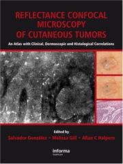
ISBN: 9780415451048 0415451043 Year: 2008 Publisher: London : Informa Healthcare,
Abstract | Keywords | Export | Availability | Bookmark
Loading...Choose an application
- Reference Manager
- EndNote
- RefWorks (Direct export to RefWorks)
Confocal microscopy --- Skin --- Microscopy, Confocal --- Skin Neoplasms --- Microscopie confocale --- Peau --- Atlases --- Cancer --- Histopathology --- Atlases. --- Atlas --- Histopathologie
ISBN: 1556426119 Year: 2002 Publisher: Thorofare : Slack Incorporated,
Abstract | Keywords | Export | Availability | Bookmark
Loading...Choose an application
- Reference Manager
- EndNote
- RefWorks (Direct export to RefWorks)
Corneal diseases --- Cornea --- Diagnostic imaging --- Microscopy, confocal --- diagnosis --- anatomy and histology --- methods
ISBN: 1872748724 9781872748726 Year: 1997 Publisher: Oxford : Bios scientific publ.,
Abstract | Keywords | Export | Availability | Bookmark
Loading...Choose an application
- Reference Manager
- EndNote
- RefWorks (Direct export to RefWorks)
Experimental solid state physics --- Optics. Quantum optics --- 681.723 --- #WSCH:MODS --- Microscopes --- 681.723 Microscopes --- CONFOCAL MICROSCOPY --- MICROSCOPY, CONFOCAL --- HANDBOOKS
Book
ISBN: 9783642219979 9783642219962 Year: 2012 Publisher: Berlin Heidelberg Springer Berlin Heidelberg Imprint Springer
Abstract | Keywords | Export | Availability | Bookmark
Loading...Choose an application
- Reference Manager
- EndNote
- RefWorks (Direct export to RefWorks)
In recent years the relevance of non-invasive bioimaging techniques in the field of melanoma screening has steadily increased. In the new era of clinicoimaging diagnosis, reflectance confocal microscopy (RCM) will have a major impact on the diagnosis and management of neoplastic and inflammatory skin diseases. This book focuses on the use and significance of in vivo RCM for non-invasive high-resolution imaging of the skin. All of the chapters in this hands-on guide are generously illustrated with numerous confocal images and structured in a reader-friendly way. The contents include: ¢ detailed information on the most relevant and up-to-date aspects of RCM, ¢ schematic drawings summarizing and explaining the most important RCM criteria, and ¢ a chapter specifically devoted to bridging the gap between dermoscopy, RCM, and histopathology. At the end of each chapter, core messages recapitulate the most pertinent aspects. Reflectance Confocal Microscopy for Skin Diseases will be a valuable resource for all physicians involved in the diagnosis and treatment of neoplastic and inflammatory skin diseases
Oncology. Neoplasms --- Paediatrics --- Otorhinolaryngology --- Pathological dermatology --- Orthopaedics. Traumatology. Plastic surgery --- Surgery --- dermatologie --- pediatrie --- plastische chirurgie --- oncologie --- chirurgie --- otorinolaryngologie --- Skin Diseases --- Microscopy, Confocal. --- Skin --- Confocal microscopy --- Peau --- Microscopie confocale --- diagnosis. --- pathology. --- Diseases --- Diagnosis. --- Maladies --- Diagnostic --- EPUB-LIV-FT LIVMEDEC SPRINGER-B

ISBN: 0471409200 9780471409205 Year: 2002 Publisher: New York Wiley-Liss
Abstract | Keywords | Export | Availability | Bookmark
Loading...Choose an application
- Reference Manager
- EndNote
- RefWorks (Direct export to RefWorks)
Biological microscopy --- Microscopy, Confocal --- Imaging, Three-Dimensional --- Microscopy, Fluorescence --- Confocal microscopy --- Fluorescence microscopy --- Three-dimensional imaging --- Microscopie confocale --- Microscopie de fluorescence --- methods --- Methodology --- Confocal microscopy. --- Fluorescence microscopy. --- Methodology. --- methods. --- 681.723 --- -#WSCH:MODS --- 3-D imaging --- 3D imaging --- Three-dimensional imaging systems --- Three-dimensional imaging techniques --- Three-dimensional visualization --- Visualization, Three-dimensional --- Imaging systems --- Microscopy --- Microscopes --- 681.723 Microscopes --- #WSCH:MODS
Book
ISBN: 0123649374 Year: 1996 Volume: 36 Publisher: San Diego ; New York ; Boston Academic Press
Abstract | Keywords | Export | Availability | Bookmark
Loading...Choose an application
- Reference Manager
- EndNote
- RefWorks (Direct export to RefWorks)
Beeldsystemen in de geneeskunde --- Experimental pathology --- Experimentele pathologie --- Geneeskunde [Beeldsystemen in de ] --- Imaging systems in medicine --- Medical imaging systems --- Medische beeldvorming --- Médecine [Système d'images en ] --- Pathologie expérimentale --- Pathology [Experimental ] --- Systèmes d'images en médecine --- Electron microscopy. --- Fluorescent dyes --- Image processing, computer-assisted --- Microscopy, confocal --- Ultrasonography
ISBN: 0896035263 159259722X 9780896035263 Year: 1999 Volume: 122 Publisher: Totowa: Humana press,
Abstract | Keywords | Export | Availability | Bookmark
Loading...Choose an application
- Reference Manager
- EndNote
- RefWorks (Direct export to RefWorks)
In Confocal Microscopy Methods and Protocols, Stephen Paddock and a highly skilled panel of experts lead the researcher using confocal techniques from the bench top, through the imaging process, to the journal page. They concisely describe all the key stages of confocal imaging-from tissue sampling methods, through the staining process, to the manipulation, presentation, and publication of the realized image. Written in a user-friendly, nontechnical style, the methods specifically cover most of the commonly used model organisms: worms, sea urchins, flies, plants, yeast, frogs, and zebrafish. Centered in the many biological applications of the confocal microscope, the book makes possible the successful imaging of both fixed and living specimens using primarily the laser scanning confocal microscope. The powerful hands-on methods collected in Confocal Microscopy Methods and Protocols will help even the novice to produce first-class cover-quality confocal images.
Confocal microscopy --- Microscopy, Confocal --- Microscopy --- Diagnostic Imaging --- Investigative Techniques --- Diagnostic Techniques and Procedures --- Analytical, Diagnostic and Therapeutic Techniques and Equipment --- Diagnosis --- Cytology --- Biology --- Health & Biological Sciences --- Confocal microscopy. --- Life sciences. --- Cell biology. --- Life Sciences. --- Cell Biology. --- Cell biology --- Cellular biology --- Cells --- Cytologists --- Biosciences --- Sciences, Life --- Science --- Microscopy. --- Analysis, Microscopic --- Light microscopy --- Micrographic analysis --- Microscope and microscopy --- Microscopic analysis --- Optical microscopy --- Optics --- Cytology.
Book
ISBN: 0387781749 0387781757 Year: 2011 Publisher: New York : Springer,
Abstract | Keywords | Export | Availability | Bookmark
Loading...Choose an application
- Reference Manager
- EndNote
- RefWorks (Direct export to RefWorks)
Most researchers agree that biological confocal microscopy was jump-started by the confocal design first published by White and Amos in 1985 in the Journal of Cell Biology. As a result, this remains a relatively young field. Yet the use of the technique has grown phenomenally since those early efforts, with new users joining the ranks daily. The publication of Basic Confocal Microscopy reflects the burgeoning need to train new students, technologists, and faculty wishing to use confocal microscopy in their research. A direct outgrowth of the authors’ five-day intensive course in the subject begun in 2005, this book covers the basics and includes all the information required to design, implement, and interpret the results of, biological experiments based on confocal microscopy. Concise yet comprehensive, the volume begins by covering the core issues of fluorescence, specimen preparation and labeling, before moving on to address the analog-to-digital conversion of specimen data gathered using confocal microscopy. Subsequent chapters detail the practicalities of operating confocal microscopes, providing all the information necessary to begin practicing confocal microscopy as well as optimizing the material obtained. The final block of chapters examine 3-dimensional analysis and the reconstruction of data sets, outline some of the ethical considerations in confocal imaging, and then supply a number of resources that the authors have found useful in their own work. Once readers have mastered the information this book presents, the resources found in its pages will be an excellent guide to continued learning about the more advanced forms of confocal microscopy.
Confocal microscopy. --- Fluorescence microscopy. --- Image processing -- Digital techniques. --- Microbiology -- Technique. --- Microscopy, Confocal. --- Microscopy, Electron, Scanning. --- Microscopy. --- Confocal microscopy --- Biology --- Health & Biological Sciences --- Microscopy --- Analysis, Microscopic --- Light microscopy --- Micrographic analysis --- Microscope and microscopy --- Microscopic analysis --- Optical microscopy --- Life sciences. --- Biotechnology. --- Cell biology. --- Life Sciences. --- Biological Microscopy. --- Cell Biology. --- Optics --- Cytology. --- Chemical engineering --- Genetic engineering --- Cell biology --- Cellular biology --- Cells --- Cytologists
| Listing 1 - 10 of 16 | << page >> |
Sort by
|