| Listing 1 - 10 of 281 | << page >> |
Sort by
|
Book
Year: 2005 Publisher: Leuven s.n.
Abstract | Keywords | Export | Availability | Bookmark
 Loading...
Choose an application
- Reference Manager
- EndNote
- RefWorks (Direct export to RefWorks)
Book
Year: 2009 Publisher: Bruxelles: UCL,
Abstract | Keywords | Export | Availability | Bookmark
 Loading...
Choose an application
- Reference Manager
- EndNote
- RefWorks (Direct export to RefWorks)
The Alzheimer’s disease (AD) is the most common form of dementia. Because of the ageing of the population, it will become a problem of public health since there is currently no treatment preventing or curing this disease. Although the exact cause of the disease is still unknown, two theories have been put forward : firstly the cholinergic hypothesis, on basis of which the cholinesterase inhibitors were developed – with a limited and only symptomatic effect – and secondly the amyloid hypothesis. It is the latter which is most largely accepted today, so that the attention is currently focused on the development of anti-amyloid therapies. In the context of studying the pathophysiology of AD, PET (Positron Emission Tomography) imaging has proved to be a valuable tool. PET-imaging is indeed a nuclear medical imaging technology which makes it possible both to quantify and to visualize in three dimensions biochemical and functional processes in living humans. This imaging technique uses common radiotracers such as [18F]-FDG (for the measurement of the glucose metabolism in the brain) and [11C]-PIB (for the visualization of amyloid deposits). [18F]-FDG is the only PET radiotracer currently used in clinical evaluations in the context of the differential diagnosis of dementia and for differentiating AD from a mild cognitive impairment, when a traditional evaluation (evaluation of cognition by neuropsychological tests, interview, clinical history…) fails. Thanks to its high diagnostic accuracy level, it allows the right selection of the patients evolving to MA with a view to their enrolment in clinical studies, avoiding erroneous conclusions and wastes of time and money linked with a bad selection of patients. Moreover, it makes it possible to evaluate the effects of new drugs by observing glucose metabolism changes. On the other hand, amyloid imaging is only used in research, but it proves to be a promising tool in the evaluation of drugs targeting amyloid as well as in the early diagnosis of the disease. PET imaging is used at all the research stages, but especially in the early stages because of its capacity to evaluate early the potential effectiveness of drugs. It prevents going on with studies on drugs which will not be appropriate. Moreover, studying the toxicity as well as the pharmacokinetics and pharmacodynamics of a drug can be performed on more restricted numbers of patients than in classical studies since the short radioactive half-life of the tracers used allows the repetition of the studies on the same subject and during the same day, eliminating any variability between the subjects. Lastly, the high sensitivity of PET makes it possible to evaluate with very low dose the distribution of the substances, thus avoiding any risk of toxicity and allowing to save several months or years compared to a traditional drug development schedule. La maladie d’Alzheimer (MA) est la forme la plus courante de démence. En raison du vieillissement de la population, elle est appelée à devenir un problème de santé publique vu qu’il n’existe actuellement aucun traitement permettant de prévenir ou de guérir cette maladie. Bien que la cause exacte de la maladie soit toujours inconnue, deux hypothèses ont été avancées : premièrement, l’hypothèse cholinergique, sur base de laquelle ont été créés les inhibiteurs de cholinestérases – dont l’effet est modeste et seulement symptomatique – et en second lieu l’hypothèse amyloïde. C’est cette dernière qui est la plus largement acceptée aujourd’hui, de sorte que l’attention se focalise actuellement sur la recherche de thérapies anti-amyloïdes. Pour comprendre la physiopathologie de la maladie, l’imagerie PET (Tomographie par Émission de Positrons) s’est avérée être un outil précieux. Le PET est en effet une méthode d’imagerie médicale nucléaire qui permet de visualiser en trois dimensions et de quantifier des processus biochimiques et fonctionnels dans le corps humain. Cette technique d’imagerie utilise des radiotraceurs dont les plus courants sont le [18F]-FDG (pour la mesure du métabolisme cérébral du glucose) et le [11C]-PIB (pour la visualisation des dépôts amyloïdes). Le [18F]-FDG est le seul radiotraceur PET qui soit actuellement utilisé en clinique, dans le cadre du diagnostic différentiel des démences et dans la distinction entre un trouble cognitif léger et la MA, lorsqu’une évaluation classique (évaluation de la cognition par des tests neuropsychologiques, interview, histoire clinique…) n’a pas abouti. Grâce à sa grande précision diagnostique, il permet la sélection judicieuse des patients évoluant vers une MA en vue de leur inclusion dans les études cliniques, évitant ainsi les conclusions erronées et les pertes de temps et d’argent dues à une mauvaise sélection de patients. De plus, il permet d’évaluer l’efficacité de nouveaux médicaments par les changements observés dans le métabolisme glucosé. L’imagerie de l’amyloïde, quant à elle, n’est encore utilisée qu’en recherche, mais elle constitue un outil prometteur dans l’évaluation des médicaments ciblant l’amyloïde ainsi que dans le diagnostic précoce de la maladie. En recherche, le PET est utilisé à toutes les étapes, mais surtout dans les stades précoces en raison de sa capacité à évaluer très tôt l’efficacité potentielle des médicaments. On évite ainsi de poursuivre l’étude de substances qui ne conviendront pas. De plus, l’étude de la toxicité, de la pharmacocinétique et de la pharmacodynamie d’un médicament peut être réalisée sur un nombre plus restreint de patients que dans les études traditionnelles. En effet, en raison de la courte demi-vie radioactive des traceurs, la répétition des études sur le même sujet et durant le même jour est possible, ce qui élimine la variabilité entre les sujets. Enfin, la haute sensibilité du PET permet d’évaluer avec de très faibles doses la distribution des substances, évitant ainsi tout risque de toxicité et faisant gagner plusieurs mois ou années par rapport à un programme classique de développement de médicament
Periodical
Abstract | Keywords | Export | Availability | Bookmark
 Loading...
Choose an application
- Reference Manager
- EndNote
- RefWorks (Direct export to RefWorks)
Book
Year: 1995 Publisher: Madrid Agencia de Evaluacion de Technologias Sanitarias (AETS)
Abstract | Keywords | Export | Availability | Bookmark
 Loading...
Choose an application
- Reference Manager
- EndNote
- RefWorks (Direct export to RefWorks)
WN 206 Tomography --- Cardiology --- Positron-Emission Tomography
Book
Year: 2009 Publisher: Bruxelles: UCL,
Abstract | Keywords | Export | Availability | Bookmark
 Loading...
Choose an application
- Reference Manager
- EndNote
- RefWorks (Direct export to RefWorks)
With the development of medical imaging techniques, care of patients with cancer is likely to be improved. If still the RECIST criteria (Response Evaluation Criteria In Solids Tumors) are used for morphological assessment of response to treatments of solid tumors, the role of functional and molecular imaging will take the extend it is more suitable for monitoring anti-tumors treatments. Indeed, from the beginning of a cytotoxic or cytostatic treatment, physicians will be available to physiological parameters of the tumors that are predictive of the effectiveness of the treatment. Biomarkers to characterize these parameters are among others: metabolic markers (18F-FDG, MRS), markers of proliferation and membrane turnover (18F-FLT, 11C-Choline and 18F-FLT), perfusion markers (DCE-MRI) and diffusion markers (DW-MRI). Moreover, these biomarkers are useful to develop new anti-tumors treatments Avec l'évolution des techniques d'imagerie médicale, la prise en charge des patients souffrant d'un cancer va vraisemblablement être améliorée. Si encore aujourd'hui les critères RECIST (Response Evaluation Criteria In Solids Tumors) sont les critères les plus utilisés pour l'évaluation morphologique de la réponse aux traitements des tumeurs solides, la place de l'imagerie fonctionnelle et moléculaire va prendre de l'ampleur puisqu'elle est bien plus adaptée pour le suivi des traitements anti-tumoraux. En effet, dès le début d'un traitement cytotoxique ou cytostatique, les médecins auront à disposition des paramètres physiologiques de la tumeur qui sont prédictifs de l'efficacité du traitement. Les marqueurs biologiques permettant de caractériser ces paramètres sont entre autres: Les marqueurs métaboliques (18F-FDG, MRS), les marqueurs de prolifération et du turnover membranaire (18F-FLT, 11C-Choline et 18F-FCH), les marqueurs de perfusion (DCE-MRI) et les marqueurs de diffusion (DW-MRI). En outre, ces biomarqueurs sont utiles pour le développement de nouveaux traitements anti-tumoraux
Positron-Emission Tomography --- Biological Markers --- Drug Therapy --- Magnetic Resonance Spectroscopy
Book
Year: 2015 Publisher: Bruxelles: UCL. Faculté de médecine et de médecine dentaire,
Abstract | Keywords | Export | Availability | Bookmark
 Loading...
Choose an application
- Reference Manager
- EndNote
- RefWorks (Direct export to RefWorks)
Objectives: the primary aim of this study is to determine the impact of Positron emission tomography PET-CT) in the management of differentiated thyroid cancer. These results will be compared the results from (PET-CT) with other imaging techniques. The second goal is to determine if there is a correlation between the SUV max and the value of thyroglobulin. Methods: forty-two patients with a histologically proven differentiated thyroid carcinoma. All patients included in this retrospective study had a PET-CT in St-Luc University Clinic between May 2007 and January 2014.We included 42 patients with an mean age of 53,5 years of whom64,3% were women. 33 patients (78,6 %) had a papillary carcinoma, 9 (21,4 %) a follicular variant of a papillary carcinoma, 7 patients (16,7%) had follicular carcinoma and 2 (4,8%) were suffering from a Hürtle cell carcinoma. In this study, 12 patients or28, 6% have a T4 as initial stage, while T2 and T3count for 11 patients each. Only 7 patients (16,7 %) had T1. When we analyze the PET-CT, 67,5% are positive 32,5% negative. Papillary carcinomas have a median SUV max of 5,6. ln contrast, the SUV max of follicular carcinomas is on average 10, 4. Some PET-CT was followed by histological examination 60% of PET-CT matched with the presence of a recurrent or metastatic lesion. In 37,8 % of cases, the scintigraphy with radioiodine is negative while the PET-CT is abnormal. An increased (unstimulated) thyroglobulin was found in 54,5 % of the patients with a positive PET-CT.Conversely, the unstimulated Tg remained normal in 20,7% of the patients with an abnormal PET-CT. In this a work, a correlation between the thyroglobulin level and he SUV max found.This correlation was more pronounced for the unstimulated thyroglobulin than with the Thyrogen-stimulated thyroglobulin. Among the positive PET-CT, a therapeutic decision was taken, of which in 16 cases a surgical treatment. Conversely, in 92% of the patients with a normal PET-CT, no therapeutic change was proposed. Conclusion : PET-CT has a important role for the detection of an aggressive recurring and/or metastatic thyroid carcinoma. It is indicated when the scintigraphy with radioiodine is negative and the thryoflobulin detectable. The high SUV max is a sign of dedifferentiated of DTC. PET-CT is important not only for the therapeutic decision but also a prognostic value. This study suggests that the PET-CT allow to adapt the treatment of DTC. Le premier but de ce travail est de déterminer l'impact de la tomographie par émission à combiner au scanner (PET-CT) dans la prise en charge du cancer différencié de la thyroïde.Mais aussi de comparer les résultats obtenus au PET-scan avec les autres techniques d'imagerie. Le second objectif sera de déterminer s'il y a une corrélation entre la SUV max et le taux de thyroglobuline.Matériel et méthodes: Quarante-deux patients présentant un carcinome différencié de la thyroïde prouvé histologiquement. Tous ces patients inclus dans cette étude rétrospective ont eu un PET-CT aux Cliniques Universitaires Saint-Luc entre mai 2007 etjanvier 2014.Résultats : Nous avons inclus 42 patients d'un âge moyen de 53,5 ans et dont 64,3% sont des femmes. 33 patient s (78,6%) présentent un carcinome papillaire, dont 9 (soit 21,4%) sont à variant folliculaire, 7 patients (16,7%) ont des carcinomes folliculaires et 2 (4,8%) étaient atteints d'un carcinome oncocytaire. Dans cette étude, les 12 patients soit 28,6% ont comme stade initial un T4, alors que le T2 et T3 comptabilisent chacun 11 patients. Seuls 7 patients soit 16,7% présentent un Tl. Lorsqu’on analyse les images PET-CT, 67,5% sont positifs et 32,5% sont négatifs. Les carcinomes papillaires ont une SUV max moyenne de 5,6. En comparaison, la SUV max moyenne des folliculaires est de 10,4. Certains PET-scan étaient suivis d'un examen histologique. 60% des PET scan concordaient avec la présence d'une lésion néoplasique à distance. Dans 37,8% des cas, la scintigraphie à l'iode radioactif est négative alors que le PET-CT est pathologique. Une Tg augmentée est détectée avec un PET-CT positif chez 54,5%. Lorsque le PET-CT est positif, on note toutefois que la thyroglobuline non stimulée peut être non détectable chez 20,7%. Dans cette étude, il existe une corrélation entre le taux de thyroglobuline et la SUV max. Mais cela est plus marqué pour la thyroglobuline non stimulée que pour celle stimulée par Thyrogen. Parmi les PET CT positifs, 37 cas soit 68,% ont bénéfic ié d'un traitement et dont 16 étaient pour un traitement chirurgical. A contrario, 92% PET-scan normaux n'entraînent aucun changement dans la thérapeutique du CDT.Conclusion :Le PET-CT a un rôle important dans la détection d'un CDT agressif récidivant et/ou métastatique. Il est donc indiqué quand la scintigraphie à l'iode radioactif est négative et la thyroglobuline détectable . La SUV max élevé est un signe de dédifférenciation du CDT. Le PET scan a donc une importance tant sur le plan thérapeutique que sur le pronostic du CDT. L'étude suggère que le PET-CT permet d'adapter la prise en charge des CDT.
Thyroid Neoplasms --- Positron-Emission Tomography --- Tomography, X-Ray Computed

ISBN: 0340816937 Year: 2005 Publisher: London : Hodder Arnold,
Abstract | Keywords | Export | Availability | Bookmark
 Loading...
Choose an application
- Reference Manager
- EndNote
- RefWorks (Direct export to RefWorks)
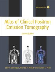
Neoplasms --- Positron-Emission Tomography --- Tomography, Emission. --- Radionuclide imaging. --- Methods.

ISBN: 0781776740 Year: 2007 Publisher: Philadelphia, PA : Lippincott Williams & Wilkins,
Abstract | Keywords | Export | Availability | Bookmark
 Loading...
Choose an application
- Reference Manager
- EndNote
- RefWorks (Direct export to RefWorks)
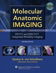
This fully updated Second Edition focuses sharply on clinical PET-CT and SPECT-CT examinations, omitting lengthy physics discussions. The book is now strictly disease oriented and integrates PET-CT and SPECT-CT applications completely. When both techniques are relevant for a disease, they are discussed together; when only one is relevant, it is discussed alone. More than 1,200 illustrations are included. A bound-in DVD contains over 80 cases to be viewed in three orthogonal planes and different CT windows organized as reference and self-assessment files. The cases provide excellent training and allow readers to test their abilities in making diagnoses on their own.
Book
ISBN: 9780781779999 Year: 2009 Publisher: Philadelphia : Lippincott Williams & Wilkins,
Abstract | Keywords | Export | Availability | Bookmark
 Loading...
Choose an application
- Reference Manager
- EndNote
- RefWorks (Direct export to RefWorks)
Positron-Emission Tomography --- Tomography, Emission. --- Tomography, X-Ray Computed --- Methods.

ISBN: 0387288368 Year: 2006 Publisher: New York : Springer,
Abstract | Keywords | Export | Availability | Bookmark
 Loading...
Choose an application
- Reference Manager
- EndNote
- RefWorks (Direct export to RefWorks)
Pediatric tomography. --- Pediatrics. --- Positron-Emission Tomography. --- Tomography, Emission.
| Listing 1 - 10 of 281 | << page >> |
Sort by
|

 Search
 Feedback
 About UniCat
  Help
Help News
News