
 Search
 Feedback
 About UniCat
  Help
Help News
News
| Listing 1 - 10 of 44 | << page >> |
Sort by
|
Book
ISBN: 0123984076 0123983983 1299773613 9780123984074 9780123983985 Year: 2013 Publisher: Burlington Elsevier Science
Abstract | Keywords | Export | Availability | Bookmark
 Loading...
Choose an application
- Reference Manager
- EndNote
- RefWorks (Direct export to RefWorks)
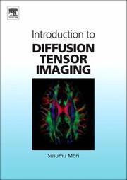
The concepts behind diffusion tensor imaging (DTI) are commonly difficult to grasp, even for magnetic resonance physicists. To make matters worse, a many more complex higher-order methods have been proposed over the last few years to overcome the now well-known deficiencies of DTI. In Introduction to Diffusion Tensor Imaging: And Higher Order Models, these concepts are explained through extensive use of illustrations rather than equations to help readers gain a more intuitive understanding of the inner workings of these techniques. Emphasis is placed on the interpretation of DTI imag
Book
Year: 2011 Publisher: Bruxelles: UCL,
Abstract | Keywords | Export | Availability | Bookmark
Loading...Choose an application
- Reference Manager
- EndNote
- RefWorks (Direct export to RefWorks)
Background: New techniques o fMRI and specially diffusion weighted MR imaging are able to perform a tractographic analysis of white matter tracts. Those technics allow to identify the position of tract close to brain lesions. Objective: The aim of the study is to identify precisely the position of a fiber tract lying near to a brain lesion, to investigate the implication of the diffusion tensor imagings tractography in the preoperative strategy and finally to evaluate this impact on the prognosis. Materials and methods: 20 patients with a tumoral or epileptic lesion underwent diffusion tensor imaging. Among these 20 patients, 3 fiber tracts were studied: the pyramidal, arcuate and visual tract. At least one of these three tracts situated near or in contact with the lesion was reconstructed in 3D. The reconstruction and the analysis of diffusion tensor data were performed using a workstation which can be viewed in 3D. 17 patients with lesions near to a fiber tract but who did flot benefit from the diffusion tensor imaging, were included as a control group. Results: The analysis of diffusion tensor data has permitted to identify the position of the fiber tract close to the lesion precisely. Tractography has resulted in a change in the surgical approach in 20%; it has redefined the extent of resection in 35% of cases and therefore had a global impact on the surgical procedure in 55% of cases. The percentage of new deficit or worsening of the pre-existing ones was 6.25% in DTI patients and 17% in the controls patients. Conclusion: Tractography by diffusion tensor imaging is a technique which can accurately identify the position of the cerebral white matter fiber tracts. It regularly influences the surgical procedure and decreased the neurological deficits at 3 months. This imaging technique provides a new perspective in understanding normal and pathological processes which affect the human brain Contexte : De nouvelles techniques d’imagerie par résonnance magnétique et plus particulièrement les séquences de diffusion permettent de réaliser une tractographie des fibres de substance blanche. Ceci permet d’identifier des faisceaux d’intérêts et des les situer par rapport à des lésions cérébrales. Objectifs: le but de l’étude est d’identifier de façon précise la position d’une lésion cérébrale par rapport au faisceau de fibre avoisinant, d’évaluer l’influence de la tractographie par imagerie du tenseur de diffusion dans la stratégie préopératoire et enfin d’estimer si ceci permet d’améliorer le pronostic. Matériels et méthodes : 20 patients avec une lésion tumorale ou épileptogène ont bénéficié de l’imagerie du tenseur de diffusion. Chez ces 20 patients, 3 faisceaux ont été étudiés: le pyramidal, l’arqué et le visuel. Au moins un de ces trois faisceaux a été reconstruit en 3D lorsqu’il(s) étai(en)t à proximité ou en contact avec la lésion. Cette reconstruction et l’analyse des données du tenseur de diffusion ont été effectuées à l’aide d’une station de travail permettant leur visualisation en 3D. 17 patients ayant une lésion dans le voisinage des faisceaux de fibres mais n’ayant pas bénéficié de l’imagerie du tenseur de diffusion constituent un groupe contrôle. Résultats : L’analyse des données du tenseur de diffusion a permis d’identifier la localisation précise des faisceaux par rapport à la lésion. La tractographie a entrainé une modification l’approche chirurgicale dans 20%, elle a redéfini l’étendue de résection dans 35% des cas et a donc eu un impact globale sur la procédure chirurgicale dans 55% des cas. Le pourcentage de nouveau déficit ou d’aggravation à 3 mois de ceux préexistants était de 6,25% chez les patients DTI et de 17% chez les coi3trôles. Conclusion : La tractographie par imagerie du tenseur de diffusion est une technique qui permet d’identifier de façon précise la position des faisceaux de substance blanche cérébrale. Elle influence de manière non négligeable la procédure chirurgicale et réduit les déficits à 3 mois. Cette technique d’imagerie offre une perspective nouvelle dans la compréhension du fonctionnement normal et des processus pathologiques qui affectent le cerveau humain
Book
Year: 2014 Publisher: Bruxelles: UCL. Faculté de médecine et de médecine dentaire,
Abstract | Keywords | Export | Availability | Bookmark
Loading...Choose an application
- Reference Manager
- EndNote
- RefWorks (Direct export to RefWorks)
Introduction : Diffusion tensor imaging (DTI) with fibertracking (FT) is a non-invasive technique able to delineate subcortical white matter tracts in 30. The purpose of this study is to analyse the impact of DTI-FT on surgical management of intra-axial tumors and especially on presurgical planning.Method : We conducted a retrospective cohort of 25 patients who underwent surgery for an intra-axial tumor near eloquent area with preoperative DTI-FT. Based on preoperati ve MR images of these patients, 6 neurosurgeons and 2 residents in neurosurgery were asked to determine their surgical strategy, first without, second with DTI-FT, on basis of 3 parameters : surgical approach, extent of resection , and predicted post-operative deficits.Results : In our study, 68% of patients underwent a gross total resection (GTR), and 33% experienced neuological worsening at 3 months after surgery. Preoperative DTI-FT had a significative impact on surgical approach (p=0,0001), on predicted extent of resection (p=0,0001), and on predicted postoperative deficits (p=0,0001). DTI-FT showed significatively more impact on presurgical planning in the group of less experienced neurosurgeons and residents, than in the group of more experienced neurosurgeons (p<0,05).Conclusion : These results indicate an impact of DTI-FT on presurgical planning, but a prospective study is needed to confirm it. Introduction : L'imagerie par tenseur de diffusion (DTI) associée à la tractographie (FT) est une technique non-invasive qui permet de reconstruire en 30 le trajet sous-cortical des faisceaux de fibres blanches cérébrales. L'objectif de cette étude est d'analyser l 'impact du DTI-FT sur la pri se en charge chirurgicale des tumeurs intra-axiales et en particulier sur le planning pré chirurgical.Méthodes : Une étude rétrospective a été menée incluant 25 patients opérés d'une tumeur intra-axiale à proximité de zones éloquentes et ayant bénéficié d'un DTI-FT pré-opératoire. A partir des images IRM pré-opératoires de ces patients, six neurochirurgiens et 2 assistants en neurochirurgie ont déterminé leur stratégie chirurgicale d'abord sans, puis avec DTI-FT, sur base de trois paramètres : la trajectoire d'abord chirurgical, l'étendue de résection tumorale, et les déficits post-opératoires permanents attendus.Résultats : Dans notre série 68% des patients ont bénéficié d'une résection macroscopiquem ent complète et 33% ont présenté une détérioration neurologique post opératoire à 3 mois. Le DTI-FT pré-opératoire a eu un impact significatif sur la trajectoire d'abord chirurgical envisagée (p=0,0001), sur l'étendue de résection prédite (p=0,0001), et sur les déficits post-opératoires permanent s annoncés (p=0,0001). Le DTI-FT a eu significativement plus d'impact sur le planning pré-chirur gical dans le groupe des neurochirurgien s et assistants de moins de dix ans d'expérience, que dans le groupe des neurochirurgien s plus expérimentés (p<0,05).Conclusion : Ces résultats plaident pour un impact du DTI-FT sur le planning pré-chirurgical , mais une étude prospective serait nécessaire pour les confirmer.
ISBN: 9780444528285 0444528288 9780080495767 0080495761 9786611013011 1281013013 Year: 2007 Publisher: Amsterdam ; Oxford : Elsevier,
Abstract | Keywords | Export | Availability | Bookmark
Loading...Choose an application
- Reference Manager
- EndNote
- RefWorks (Direct export to RefWorks)
The concept of Diffusion Tensor Imaging (DTI) is often difficult to grasp, even for Magnetic Resonance physicists. Introduction to Diffusion Tensor Imaging uses extensive illustrations (not equations) to help readers to understand how DTI works. Emphasis is placed on the interpretation of DTI images, the design of DTI experiments, and the forms of application studies. The theory of DTI is constantly evolving and so there is a need for a textbook that explains how the technique works in a way that is easy to understand - Introduction to Diffusion Tensor Imaging fills this gap.
Spectrometric and optical chemical analysis --- Physical methods for diagnosis --- fysicochemie --- Diffusion tensor imaging. --- Brain --- DTI (Diffusion tensor imaging) --- Magnetic resonance diffusion tensor imaging --- Diffusion magnetic resonance imaging --- Magnetic resonance imaging.
Multi
ISBN: 9780123983985 9780123984074 0123984076 0123983983 Year: 2014 Publisher: Oxford, England ; San Diego, Calif. : Academic Press,
Abstract | Keywords | Export | Availability | Bookmark
Loading...Choose an application
- Reference Manager
- EndNote
- RefWorks (Direct export to RefWorks)
The concepts behind diffusion tensor imaging (DTI) are commonly difficult to grasp, even for magnetic resonance physicists. To make matters worse, a many more complex higher-order methods have been proposed over the last few years to overcome the now well-known deficiencies of DTI. In Introduction to Diffusion Tensor Imaging: And Higher Order Models, these concepts are explained through extensive use of illustrations rather than equations to help readers gain a more intuitive understanding of the inner workings of these techniques. Emphasis is placed on the interpretation of DTI imag
Physical methods for diagnosis --- Brain --- Diffusion tensor imaging. --- Magnetic resonance imaging.
Book
Abstract | Keywords | Export | Availability | Bookmark
Loading...Choose an application
- Reference Manager
- EndNote
- RefWorks (Direct export to RefWorks)
This e-book focuses primarily on the role of the fornix as a functional, prognostic, and diagnostic marker of Alzheimer’s disease (AD), and the application of such a marker in clinical practice. Researchers have long been focused on the cortical pathology of AD, since the most important pathologic features are the senile plaques found in the cortex, and the neurofibrillary tangles and neuronal loss that start from the entorhinal cortex and the hippocampus. In addition to gray matter structures, histopathological studies indicate that the white matter is also altered in AD. The fornix is a white matter bundle that constitutes a core element of the limbic circuits, and is one of the most important anatomical structures related to memory. The fornices originate from the bilateral hippocampi, merge at the midline of the brain, again divide into the left and right side, and then into the precommissural and the postcommissural fibers, and terminate at the septal nuclei, nucleus accumbens (precommissural fornix), and hypothalamus (postcommissural fornix). These functional and anatomical features of the fornix have naturally captured researchers’ attention as possible diagnostic and prognostic markers of AD. Growing evidence indicates that the alterations seen in the fornix are potentially a good marker with which to predict future conversion from mild cognitive impairment to AD, and even from a cognitively normal state to AD. The degree of alteration is correlated with the degree of memory impairment, indicating the potential for the use of the fornix as a functional marker. Moreover, there have been attempts to stimulate the fornix to recover the cognitive function lost with AD. Our goal is to provide information about the status of current research and to facilitate further scientific and clinical advancement in this topic.
Fornix --- Limbic --- Memory --- normal aging --- Cognition --- Mild Cognitive Impairment --- Alzheimer's disease --- Diffusion Tensor Imaging
Book
Abstract | Keywords | Export | Availability | Bookmark
Loading...Choose an application
- Reference Manager
- EndNote
- RefWorks (Direct export to RefWorks)
This e-book focuses primarily on the role of the fornix as a functional, prognostic, and diagnostic marker of Alzheimer’s disease (AD), and the application of such a marker in clinical practice. Researchers have long been focused on the cortical pathology of AD, since the most important pathologic features are the senile plaques found in the cortex, and the neurofibrillary tangles and neuronal loss that start from the entorhinal cortex and the hippocampus. In addition to gray matter structures, histopathological studies indicate that the white matter is also altered in AD. The fornix is a white matter bundle that constitutes a core element of the limbic circuits, and is one of the most important anatomical structures related to memory. The fornices originate from the bilateral hippocampi, merge at the midline of the brain, again divide into the left and right side, and then into the precommissural and the postcommissural fibers, and terminate at the septal nuclei, nucleus accumbens (precommissural fornix), and hypothalamus (postcommissural fornix). These functional and anatomical features of the fornix have naturally captured researchers’ attention as possible diagnostic and prognostic markers of AD. Growing evidence indicates that the alterations seen in the fornix are potentially a good marker with which to predict future conversion from mild cognitive impairment to AD, and even from a cognitively normal state to AD. The degree of alteration is correlated with the degree of memory impairment, indicating the potential for the use of the fornix as a functional marker. Moreover, there have been attempts to stimulate the fornix to recover the cognitive function lost with AD. Our goal is to provide information about the status of current research and to facilitate further scientific and clinical advancement in this topic.
Fornix --- Limbic --- Memory --- normal aging --- Cognition --- Mild Cognitive Impairment --- Alzheimer's disease --- Diffusion Tensor Imaging --- Fornix --- Limbic --- Memory --- normal aging --- Cognition --- Mild Cognitive Impairment --- Alzheimer's disease --- Diffusion Tensor Imaging
Book
ISBN: 1493931180 1493931172 9781493931170 Year: 2016 Publisher: New York, NY : Springer New York : Imprint: Springer,
Abstract | Keywords | Export | Availability | Bookmark
Loading...Choose an application
- Reference Manager
- EndNote
- RefWorks (Direct export to RefWorks)
This book provides an overview of the practical aspects of diffusion tensor imaging (DTI), from understanding the basis of the technique through selection of the right protocols, trouble-shooting data quality, and analyzing DTI data optimally. DTI is a non-invasive magnetic resonance imaging (MRI) technique for visualizing and quantifying tissue microstructure based on diffusion. The book discusses the theoretical background underlying DTI and advanced techniques based on higher-order models and multi-shell diffusion imaging. It covers the practical implementation of DTI; derivation of information from DTI data; and a range of clinical applications, including neurosurgical planning and the assessment of brain tumors. Its practical utility is enhanced by decision schemes and a fully annotated DTI brain atlas, including color fractional anisotropy maps and 3D tractography reconstructions of major white matter fiber bundles. Featuring contributions from leading specialists in the field of DTI, Diffusion Tensor Imaging: A Practical Handbook is a valuable resource for radiologists, neuroradiologists, MRI technicians, and clinicians.
Brain --- Diffusion tensor imaging. --- Magnetic resonance imaging. --- DTI (Diffusion tensor imaging) --- Magnetic resonance diffusion tensor imaging --- Diffusion magnetic resonance imaging --- Radiology, Medical. --- Neurology. --- Neurosciences. --- Neuroradiology. --- Diagnostic Radiology. --- Neural sciences --- Neurological sciences --- Neuroscience --- Medical sciences --- Nervous system --- Medicine --- Neuropsychiatry --- Clinical radiology --- Radiology, Medical --- Radiology (Medicine) --- Medical physics --- Diseases --- Diffusion Tensor Imaging --- DTI MRI --- Diffusion Tensor MRI --- Diffusion Tensor Magnetic Resonance Imaging --- Diffusion Tractography --- Diffusion Tensor MRIs --- Imaging, Diffusion Tensor --- MRI, Diffusion Tensor --- Tractography, Diffusion --- Radiology. --- Neurology . --- Radiological physics --- Physics --- Radiation --- Neuroradiography --- Neuroradiology
Book
ISBN: 3642204554 3642204562 1299336523 Year: 2013 Publisher: Berlin : Springer,
Abstract | Keywords | Export | Availability | Bookmark
Loading...Choose an application
- Reference Manager
- EndNote
- RefWorks (Direct export to RefWorks)
Diffusion Tensor Imaging (DTI) is a variation of diffusion-weighed imaging. Particularly in the neurosciences, this technique has gained tremendous momentum in the past decade, both from a technical point of view as well as in its applications. DTI is mainly used in neurological diagnosis and psychiatric and neurologic research, e.g. in order to locate brain tumors and depict their invasivity. DTI offers a unique in-vivo insight into the three-dimensional structure of the human central nervous system. While easy interpretation and evaluation is often hampered by the complexity of both the technique and neuroanatomy, this atlas helps you recognize every one of the important structures rapidly and unambiguously. In the introduction, this atlas provides a concise outline of the evolution of diffusion imaging and describes its potential applications. In the core part of the atlas, the neuroanatomically important structures are clearly labeled both on DTI-derived color maps and conventional MRI. Complex fiber architecture is illustrated schematically and described concisely in textboxes directly on the relevant page. In the final part of the atlas, a straightforward, step-by-step approach for the three-dimensional reconstruction of the most prominent fiber structures is given, and potential pitfalls are indicated. The atlas aims at neuroscientists, neuroanatomists, neurologists, psychiatrists, clinical psychologists, physicists, and computer scientists. For advanced users, the atlas may serve as a reference work, while students and scientists are thoroughly introduced into DTI.
Brain -- Atlases. --- Brain -- Magnetic resonance imaging. --- Brain. --- Diffusion tensor imaging. --- Brain --- Diffusion tensor imaging --- Diagnostic Techniques, Neurological --- Publication Formats --- Investigative Techniques --- Diffusion Magnetic Resonance Imaging --- Publication Characteristics --- Analytical, Diagnostic and Therapeutic Techniques and Equipment --- Magnetic Resonance Imaging --- Diagnostic Techniques and Procedures --- Diagnosis --- Diagnostic Imaging --- Atlases --- Diffusion Tensor Imaging --- Medicine --- Health & Biological Sciences --- Neurology --- Magnetic resonance imaging --- DTI (Diffusion tensor imaging) --- Magnetic resonance diffusion tensor imaging --- Medicine. --- Radiology. --- Neurology. --- Psychiatry. --- Medicine & Public Health. --- Imaging / Radiology. --- Diffusion magnetic resonance imaging --- Radiology, Medical. --- Medicine and psychology --- Mental health --- Psychology, Pathological --- Clinical radiology --- Radiology, Medical --- Radiology (Medicine) --- Medical physics --- Nervous system --- Neuropsychiatry --- Diseases --- Neurology . --- Radiological physics --- Physics --- Radiation
| Listing 1 - 10 of 44 | << page >> |
Sort by
|