| Listing 1 - 10 of 12 | << page >> |
Sort by
|
Book
Year: 2008 Publisher: Rockville, MD : U.S. Dept. of Health and Human Services, Food and Drug Administration, Office of Combination Products (OCP) in Office of Commissioner : Center for Devices and Radiological Health (CDRH) : Center for Drug Evaluation and Research (CDER),
Abstract | Keywords | Export | Availability | Bookmark
 Loading...
Choose an application
- Reference Manager
- EndNote
- RefWorks (Direct export to RefWorks)
Contrast-enhanced magnetic resonance imaging --- Contrast-enhanced ultrasound --- Biomedical materials --- Equipment and supplies --- Evaluation. --- Equipment and supplies --- Evaluation. --- Evaluation.
Book
Year: 2008 Publisher: Rockville, MD : U.S. Dept. of Health and Human Services, Food and Drug Administration, Office of Combination Products (OCP) in Office of Commissioner : Center for Devices and Radiological Health (CDRH) : Center for Drug Evaluation and Research (CDER),
Abstract | Keywords | Export | Availability | Bookmark
 Loading...
Choose an application
- Reference Manager
- EndNote
- RefWorks (Direct export to RefWorks)
Contrast-enhanced magnetic resonance imaging --- Contrast-enhanced ultrasound --- Biomedical materials --- Equipment and supplies --- Evaluation. --- Equipment and supplies --- Evaluation. --- Evaluation.

ISBN: 3540589759 Year: 1996 Publisher: Berlin : Springer,
Abstract | Keywords | Export | Availability | Bookmark
 Loading...
Choose an application
- Reference Manager
- EndNote
- RefWorks (Direct export to RefWorks)
Breast Diseases --- Breast Implants. --- Breast Neoplasms --- Breast --- Contrast Media. --- Contrast-enhanced magnetic resonance imaging. --- Image Enhancement. --- Magnetic Resonance Imaging. --- diagnosis. --- diagnosis. --- Magnetic resonance imaging.
Book
ISBN: 9781119991762 Year: 2013 Publisher: Hoboken, NJ : John Wiley & Sons Inc.,
Abstract | Keywords | Export | Availability | Bookmark
 Loading...
Choose an application
- Reference Manager
- EndNote
- RefWorks (Direct export to RefWorks)
"The second edition of The Chemistry of Contrast Agents in Medical Magnetic Resonance Imaging is a comprehensive treaty covering all aspects of production, use, operating mechanism and theory of these diagnostic agents used to produce high contrast images in MRI..It has been completed to now include recent developments on "classical" Gd-based and iron-oxide probes and chapters dedicated to the most significant advances in molecular imaging probes. Chemical Exchange Saturation Transfer is discussed, which is a novel means of generating MRI contrast"-- "This treatise covers all aspects of production, use, operating mechanism and theory of these diagnostic agents used to produce high contrast images in MRI"--

ISBN: 0813159067 9780813159065 9780813119441 0813119448 9780813119441 Year: 1997 Publisher: Lexington, Kentucky : The University Press of Kentucky,
Abstract | Keywords | Export | Availability | Bookmark
 Loading...
Choose an application
- Reference Manager
- EndNote
- RefWorks (Direct export to RefWorks)
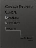
In Contrast-Enhanced Clinical Magnetic Resonance Imaging, Val M. Runge and other leading experts present an overview of the basic principles regarding MR contrast media, a review of clinical applications in the head, spine, and body, and a look at future developments. Their focus is on clinical applications, with extensive illustrations to demonstrate the use of MR in each anatomic area and to aid in film interpretation.
Contrast-enhanced magnetic resonance imaging. --- Magnetic resonance imaging. --- Contrast-enhanced MRI --- Magnetic resonance imaging --- Clinical magnetic resonance imaging --- Diagnostic magnetic resonance imaging --- Functional magnetic resonance imaging --- Imaging, Magnetic resonance --- Medical magnetic resonance imaging --- MR imaging --- MRI (Magnetic resonance imaging) --- NMR imaging --- Nuclear magnetic resonance --- Nuclear magnetic resonance imaging --- Cross-sectional imaging --- Diagnostic imaging --- Diagnostic use
Book
ISBN: 8847056756 8847056705 1322174938 Year: 2014 Publisher: Milano : Springer Milan : Imprint: Springer,
Abstract | Keywords | Export | Availability | Bookmark
 Loading...
Choose an application
- Reference Manager
- EndNote
- RefWorks (Direct export to RefWorks)
In recent years, there has been huge interest in developing new methods that offer improved accuracy for the detection of small bowel pathology, and in particular for the assessment of inflammatory bowel diseases (IBD). Cross-sectional imaging, such as CT and MR, has advantages over traditional barium fluoroscopic techniques in terms of direct visualization of the bowel wall and improved visualization of extraluminal findings and complications. This means a complete change in the diagnostic approach to the patient with IBD: from analysis of the bowel surface to direct evaluation of parietal alterations and assessment of peri- and extraintestinal involvement. The ideal imaging test is reproducible, well tolerated by patients and, above all, free of ionizing radiation. MR enterography, currently performed only in a few reference centers, meets these criteria and offers accurate diagnosis, particularly in respect of the wide spectrum of intra- and extraintestinal complications of IBD. This book provides a thorough overview of the indications, techniques, diagnostic advantages, and limitations of MR enterography. Particular attention is paid to patient preparation in relation to the particular study type and to the potential advantages of the most up-to-date MR studies in specific cases, e.g., allergy or renal failure. A separate chapter is devoted to MR of perianal region for the detection and staging of perianal fistula, a common complication in patients with Crohn’s disease. Numerous high-quality illustrations are included and help to ensure that the book will be a valuable source of information for every radiologist involved in abdominal MR imaging.
Contrast-enhanced magnetic resonance imaging. --- Inflammatory bowel diseases --- Magnetic resonance imaging. --- IBD (Disease) --- Inflammatory bowel disease --- Intestines --- Gastroenteritis --- Contrast-enhanced MRI --- Magnetic resonance imaging --- Inflammation --- Radiology, Medical. --- Gastroenterology. --- Pediatrics. --- Endocrinology. --- Abdomen --- Imaging / Radiology. --- Diagnostic Radiology. --- Proctology. --- Abdominal Surgery. --- Surgery. --- Abdominal surgery --- Laparotomy --- Internal medicine --- Hormones --- Paediatrics --- Pediatric medicine --- Medicine --- Children --- Digestive organs --- Clinical radiology --- Radiology, Medical --- Radiology (Medicine) --- Medical physics --- Diseases --- Health and hygiene --- Radiology. --- Gastroenterology . --- Abdominal surgery. --- Gastroenterology --- Radiological physics --- Physics --- Radiation
Book

ISBN: 3319646389 3319646370 Year: 2018 Publisher: Cham : Springer International Publishing : Imprint: Springer,
Abstract | Keywords | Export | Availability | Bookmark
 Loading...
Choose an application
- Reference Manager
- EndNote
- RefWorks (Direct export to RefWorks)
This book provides a comprehensive survey of the pharmacokinetic models used for the quantitative interpretation of contrast-enhanced imaging. It discusses all the available imaging technologies and the problems related to the calibration of the imaging system and accuracy of the estimated physiological parameters. Enhancing imaging modalities using contrast agents has opened up new opportunities for going beyond morphological information and enabling minimally invasive assessment of tissue and organ functionality down to the molecular level. In combination with mathematical modeling of the contrast agent kinetics, contrast- enhanced imaging has the potential to provide clinically valuable additional information by estimating quantitative physiological parameters. The book presents the broad spectrum of diagnostic possibilities provided by quantitative contrast-enhanced imaging, with a particular focus on cardiology and oncology, as well as novel developments in the area of quantitative molecular imaging along with their potential clinical applications. Given the variety of available techniques, the choice of the appropriate imaging modality and the most suitable pharmacokinetic model is often challenging. As such, the book provides a valuable technical guide for researchers, clinical scientists, and experts in the field who wish to better understand and properly apply tracer-kinetic modeling for quantitative contrast-enhanced imaging.
Contrast-enhanced magnetic resonance imaging. --- Contrast media (Diagnostic imaging) --- Contrast agents (Diagnostic imaging) --- Contrast media --- Diagnostic imaging --- Contrast-enhanced MRI --- Magnetic resonance imaging --- Equipment and supplies --- Radiology, Medical. --- Oncology. --- Imaging / Radiology. --- Signal, Image and Speech Processing. --- Cancer Research. --- Biological and Medical Physics, Biophysics. --- Tumors --- Clinical radiology --- Radiology, Medical --- Radiology (Medicine) --- Medical physics --- Radiology. --- Signal processing. --- Image processing. --- Speech processing systems. --- Cancer research. --- Biophysics. --- Biological physics. --- Biological physics --- Biology --- Medical sciences --- Physics --- Cancer research --- Computational linguistics --- Electronic systems --- Information theory --- Modulation theory --- Oral communication --- Speech --- Telecommunication --- Singing voice synthesizers --- Pictorial data processing --- Picture processing --- Processing, Image --- Imaging systems --- Optical data processing --- Processing, Signal --- Information measurement --- Signal theory (Telecommunication) --- Radiological physics --- Radiation
Book
ISBN: 3642097154 3540784225 9786612128011 1282128019 3540784233 Year: 2009 Publisher: Berlin : Springer,
Abstract | Keywords | Export | Availability | Bookmark
 Loading...
Choose an application
- Reference Manager
- EndNote
- RefWorks (Direct export to RefWorks)
MRI Atlas of Prostate Cancer analyses high-resolution MRI scanning and dynamic contrast-enhanced (DCE) MRI. This combination improves the diagnosis and staging of prostate cancer and may replace PSA testing and digital rectal examination within the next decade. The book commences with chapters on normal anatomy, anatomic variations, benign disease and intraprostatic tumors. The subsequent chapters on MRI of extracapsular disease create a useful atlas of pathologic anatomy. This is the first text of its kind to show color-coded DCE-MRI scans of prostate cancer and to correlate these imaging findings with tumor grading. The chapters on the post-treatment prostate clearly display the increasing incidence of post-therapy recurrences and note alternative treatment options. A further chapter containing a number of case studies familiarizes the reader with the most common findings as well as the pitfalls that oncologists face in the management of prostate cancer. This book is intended for internists, radiologists, radiotherapists, oncologists, urologists, family practitioners, and general surgeons. Ultrasound, MRI, and radiotherapy technicians will find it extremely useful as a reference guide.
Atlases, Pictorial. --- Contrast Media. --- Contrast-enhanced magnetic resonance imaging. --- Magnetic resonance imaging. --- Prostate -- Cancer -- Magnetic resonance imaging. --- Prostatic Neoplasms -- diagnosis. --- Genital Neoplasms, Male --- Specialty Uses of Chemicals --- Diagnostic Imaging --- Tomography --- Prostatic Diseases --- Chemical Actions and Uses --- Urogenital Neoplasms --- Genital Diseases, Male --- Diagnostic Techniques and Procedures --- Male Urogenital Diseases --- Diagnosis --- Neoplasms by Site --- Chemicals and Drugs --- Diseases --- Neoplasms --- Analytical, Diagnostic and Therapeutic Techniques and Equipment --- Prostatic Neoplasms --- Magnetic Resonance Imaging --- Contrast Media --- Medicine --- Health & Biological Sciences --- Radiology, MRI, Ultrasonography & Medical Physics --- Oncology --- Prostate --- Cancer --- Gland, Prostate --- Glandula prostata --- Prostata --- Prostate gland --- Contrast-enhanced MRI --- Medicine. --- Radiology. --- Oncology. --- Urology. --- Medicine & Public Health. --- Imaging / Radiology. --- Exocrine glands --- Generative organs, Male --- Magnetic resonance imaging --- Radiology, Medical. --- Oncology . --- Tumors --- Genitourinary organs --- Clinical radiology --- Radiology, Medical --- Radiology (Medicine) --- Medical physics --- Radiological physics --- Physics --- Radiation
Book
Year: 2021 Publisher: Basel, Switzerland MDPI - Multidisciplinary Digital Publishing Institute
Abstract | Keywords | Export | Availability | Bookmark
 Loading...
Choose an application
- Reference Manager
- EndNote
- RefWorks (Direct export to RefWorks)
undefined
Medicine. --- positron emission tomography --- head and neck neoplasms --- neovascularization --- pathologic --- PET/CT --- urothelial carcinoma --- bladder cancer --- upper tract urothelial carcinoma --- survival --- PET --- PSMA --- prostate --- DCFPyL --- DCFBC --- PSMA-1007 --- ovarian cancer --- relapse --- SUVmax --- targeted therapy --- prognosis --- soft tissue sarcoma (STS) --- pazopanib --- dynamic 18F-FDG PET/CT --- SUV --- two-tissue compartment model --- magnetic resonance imaging --- machine learning --- diffusion --- perfusion --- texture analysis --- squamous cell carcinoma of the head and neck --- diffusion-weighted imaging --- malignant pleural mesothelioma --- pleural dissemination --- empyema --- pleural effusion --- mCRPC --- SPECT/CT --- Computer-assisted diagnosis --- XOFIGO --- Therapy response assessment --- circulating miRNAs --- breast cancer --- imaging parameters --- PET/MRI --- biomarkers --- triple negative breast cancer --- VCAM-1 --- SPECT imaging --- sdAbs --- Hounsfield unit --- computed tomography --- adipose tissue --- precision oncology --- FDG-PET/CT --- PERCIST --- metastatic breast cancer --- prostate cancer --- 18F-FACBC --- recurrence --- meta-analysis --- review --- meningioma --- somatostatin receptor --- neuroimaging --- radionuclide therapy --- breast --- imaging --- marker --- radiomics --- Yin Yang 1 --- PDAC --- Mesothelin --- noninvasive imaging --- receptor status --- molecular imaging --- nuclear medicine --- guidelines --- overutilization --- epistemology --- consensus --- mantle cell lymphoma --- 18F-FDG PET/CT --- Deauville criteria --- Radium-223 --- FDG --- castrate resistant prostate cancer --- programmed cell death 1 receptor --- diagnostic imaging --- CTLA-4 Antigen --- Immunotherapy --- Adoptive --- radioactive tracers --- radionuclide imaging --- CD8-Positive T-Lymphocytes --- PI-RADS --- diffusion kurtosis imaging --- dynamic contrast-enhanced magnetic resonance imaging --- 68Gallium-PSMA PET/CT --- prostate-specific-antigen --- PSA kinetics thresholds --- biochemical recurrence --- optimal cutoff level --- non-small-cell lung cancer --- circulating tumor cells --- immunotherapy --- response to treatment --- head and neck cancer --- HPV --- EBV --- p16 --- Molecular imaging --- miRNA expression --- radiogenomics --- radiomic --- diagnosis --- biomarker --- glioblastoma --- radiation therapy --- MRI --- diffusion tensor imaging --- Hodgkin lymphoma --- diffuse large B-cell lymphoma --- staging --- response assessment --- locally advanced cervical cancer --- concurrent chemoradiotherapy --- treatment response --- follow up --- cystic tumor --- International Consensus Guidelines --- intraductal papillary mucinous neoplasms --- pancreatic neoplasms --- PD-1 --- PD-L1 --- response to therapy --- NSCLC --- positron-emission tomography --- single-photon emission computed tomography --- immune checkpoint inhibitors --- gold nanoparticle --- heat shock protein 70 --- spectral-CT --- positron emission tomography --- head and neck neoplasms --- neovascularization --- pathologic --- PET/CT --- urothelial carcinoma --- bladder cancer --- upper tract urothelial carcinoma --- survival --- PET --- PSMA --- prostate --- DCFPyL --- DCFBC --- PSMA-1007 --- ovarian cancer --- relapse --- SUVmax --- targeted therapy --- prognosis --- soft tissue sarcoma (STS) --- pazopanib --- dynamic 18F-FDG PET/CT --- SUV --- two-tissue compartment model --- magnetic resonance imaging --- machine learning --- diffusion --- perfusion --- texture analysis --- squamous cell carcinoma of the head and neck --- diffusion-weighted imaging --- malignant pleural mesothelioma --- pleural dissemination --- empyema --- pleural effusion --- mCRPC --- SPECT/CT --- Computer-assisted diagnosis --- XOFIGO --- Therapy response assessment --- circulating miRNAs --- breast cancer --- imaging parameters --- PET/MRI --- biomarkers --- triple negative breast cancer --- VCAM-1 --- SPECT imaging --- sdAbs --- Hounsfield unit --- computed tomography --- adipose tissue --- precision oncology --- FDG-PET/CT --- PERCIST --- metastatic breast cancer --- prostate cancer --- 18F-FACBC --- recurrence --- meta-analysis --- review --- meningioma --- somatostatin receptor --- neuroimaging --- radionuclide therapy --- breast --- imaging --- marker --- radiomics --- Yin Yang 1 --- PDAC --- Mesothelin --- noninvasive imaging --- receptor status --- molecular imaging --- nuclear medicine --- guidelines --- overutilization --- epistemology --- consensus --- mantle cell lymphoma --- 18F-FDG PET/CT --- Deauville criteria --- Radium-223 --- FDG --- castrate resistant prostate cancer --- programmed cell death 1 receptor --- diagnostic imaging --- CTLA-4 Antigen --- Immunotherapy --- Adoptive --- radioactive tracers --- radionuclide imaging --- CD8-Positive T-Lymphocytes --- PI-RADS --- diffusion kurtosis imaging --- dynamic contrast-enhanced magnetic resonance imaging --- 68Gallium-PSMA PET/CT --- prostate-specific-antigen --- PSA kinetics thresholds --- biochemical recurrence --- optimal cutoff level --- non-small-cell lung cancer --- circulating tumor cells --- immunotherapy --- response to treatment --- head and neck cancer --- HPV --- EBV --- p16 --- Molecular imaging --- miRNA expression --- radiogenomics --- radiomic --- diagnosis --- biomarker --- glioblastoma --- radiation therapy --- MRI --- diffusion tensor imaging --- Hodgkin lymphoma --- diffuse large B-cell lymphoma --- staging --- response assessment --- locally advanced cervical cancer --- concurrent chemoradiotherapy --- treatment response --- follow up --- cystic tumor --- International Consensus Guidelines --- intraductal papillary mucinous neoplasms --- pancreatic neoplasms --- PD-1 --- PD-L1 --- response to therapy --- NSCLC --- positron-emission tomography --- single-photon emission computed tomography --- immune checkpoint inhibitors --- gold nanoparticle --- heat shock protein 70 --- spectral-CT
Book
Year: 2021 Publisher: Basel, Switzerland MDPI - Multidisciplinary Digital Publishing Institute
Abstract | Keywords | Export | Availability | Bookmark
 Loading...
Choose an application
- Reference Manager
- EndNote
- RefWorks (Direct export to RefWorks)
The issue of Cancers Journal entitled “Role of Medical Imaging in Cancers” presents a detailed summary of evidences about molecular imaging, including the role of computed tomography (CT), magnetic resonance imaging (MRI), single photon emission tomography (SPET) and positron emission tomography (PET) or PET/CT or PET/MR imaging in many type of tumors (i.e. sarcoma, prostate, breast and others), motivating the role of these imaging modalities in different setting of disease and showing the recent developments, in terms of radiopharmaceuticals, software and artificial intelligence in this field. The collection of articles is very useful for many specialists, because it has been conceived for a multidisciplinary point of view, in order to drive to a personalized medicine.
Medicine. --- positron emission tomography --- head and neck neoplasms --- neovascularization --- pathologic --- PET/CT --- urothelial carcinoma --- bladder cancer --- upper tract urothelial carcinoma --- survival --- PET --- PSMA --- prostate --- DCFPyL --- DCFBC --- PSMA-1007 --- ovarian cancer --- relapse --- SUVmax --- targeted therapy --- prognosis --- soft tissue sarcoma (STS) --- pazopanib --- dynamic 18F-FDG PET/CT --- SUV --- two-tissue compartment model --- magnetic resonance imaging --- machine learning --- diffusion --- perfusion --- texture analysis --- squamous cell carcinoma of the head and neck --- diffusion-weighted imaging --- malignant pleural mesothelioma --- pleural dissemination --- empyema --- pleural effusion --- mCRPC --- SPECT/CT --- Computer-assisted diagnosis --- XOFIGO --- Therapy response assessment --- circulating miRNAs --- breast cancer --- imaging parameters --- PET/MRI --- biomarkers --- triple negative breast cancer --- VCAM-1 --- SPECT imaging --- sdAbs --- Hounsfield unit --- computed tomography --- adipose tissue --- precision oncology --- FDG-PET/CT --- PERCIST --- metastatic breast cancer --- prostate cancer --- 18F-FACBC --- recurrence --- meta-analysis --- review --- meningioma --- somatostatin receptor --- neuroimaging --- radionuclide therapy --- breast --- imaging --- marker --- radiomics --- Yin Yang 1 --- PDAC --- Mesothelin --- noninvasive imaging --- receptor status --- molecular imaging --- nuclear medicine --- guidelines --- overutilization --- epistemology --- consensus --- mantle cell lymphoma --- 18F-FDG PET/CT --- Deauville criteria --- Radium-223 --- FDG --- castrate resistant prostate cancer --- programmed cell death 1 receptor --- diagnostic imaging --- CTLA-4 Antigen --- Immunotherapy --- Adoptive --- radioactive tracers --- radionuclide imaging --- CD8-Positive T-Lymphocytes --- PI-RADS --- diffusion kurtosis imaging --- dynamic contrast-enhanced magnetic resonance imaging --- 68Gallium-PSMA PET/CT --- prostate-specific-antigen --- PSA kinetics thresholds --- biochemical recurrence --- optimal cutoff level --- non-small-cell lung cancer --- circulating tumor cells --- immunotherapy --- response to treatment --- head and neck cancer --- HPV --- EBV --- p16 --- Molecular imaging --- miRNA expression --- radiogenomics --- radiomic --- diagnosis --- biomarker --- glioblastoma --- radiation therapy --- MRI --- diffusion tensor imaging --- Hodgkin lymphoma --- diffuse large B-cell lymphoma --- staging --- response assessment --- locally advanced cervical cancer --- concurrent chemoradiotherapy --- treatment response --- follow up --- cystic tumor --- International Consensus Guidelines --- intraductal papillary mucinous neoplasms --- pancreatic neoplasms --- PD-1 --- PD-L1 --- response to therapy --- NSCLC --- positron-emission tomography --- single-photon emission computed tomography --- immune checkpoint inhibitors --- gold nanoparticle --- heat shock protein 70 --- spectral-CT --- positron emission tomography --- head and neck neoplasms --- neovascularization --- pathologic --- PET/CT --- urothelial carcinoma --- bladder cancer --- upper tract urothelial carcinoma --- survival --- PET --- PSMA --- prostate --- DCFPyL --- DCFBC --- PSMA-1007 --- ovarian cancer --- relapse --- SUVmax --- targeted therapy --- prognosis --- soft tissue sarcoma (STS) --- pazopanib --- dynamic 18F-FDG PET/CT --- SUV --- two-tissue compartment model --- magnetic resonance imaging --- machine learning --- diffusion --- perfusion --- texture analysis --- squamous cell carcinoma of the head and neck --- diffusion-weighted imaging --- malignant pleural mesothelioma --- pleural dissemination --- empyema --- pleural effusion --- mCRPC --- SPECT/CT --- Computer-assisted diagnosis --- XOFIGO --- Therapy response assessment --- circulating miRNAs --- breast cancer --- imaging parameters --- PET/MRI --- biomarkers --- triple negative breast cancer --- VCAM-1 --- SPECT imaging --- sdAbs --- Hounsfield unit --- computed tomography --- adipose tissue --- precision oncology --- FDG-PET/CT --- PERCIST --- metastatic breast cancer --- prostate cancer --- 18F-FACBC --- recurrence --- meta-analysis --- review --- meningioma --- somatostatin receptor --- neuroimaging --- radionuclide therapy --- breast --- imaging --- marker --- radiomics --- Yin Yang 1 --- PDAC --- Mesothelin --- noninvasive imaging --- receptor status --- molecular imaging --- nuclear medicine --- guidelines --- overutilization --- epistemology --- consensus --- mantle cell lymphoma --- 18F-FDG PET/CT --- Deauville criteria --- Radium-223 --- FDG --- castrate resistant prostate cancer --- programmed cell death 1 receptor --- diagnostic imaging --- CTLA-4 Antigen --- Immunotherapy --- Adoptive --- radioactive tracers --- radionuclide imaging --- CD8-Positive T-Lymphocytes --- PI-RADS --- diffusion kurtosis imaging --- dynamic contrast-enhanced magnetic resonance imaging --- 68Gallium-PSMA PET/CT --- prostate-specific-antigen --- PSA kinetics thresholds --- biochemical recurrence --- optimal cutoff level --- non-small-cell lung cancer --- circulating tumor cells --- immunotherapy --- response to treatment --- head and neck cancer --- HPV --- EBV --- p16 --- Molecular imaging --- miRNA expression --- radiogenomics --- radiomic --- diagnosis --- biomarker --- glioblastoma --- radiation therapy --- MRI --- diffusion tensor imaging --- Hodgkin lymphoma --- diffuse large B-cell lymphoma --- staging --- response assessment --- locally advanced cervical cancer --- concurrent chemoradiotherapy --- treatment response --- follow up --- cystic tumor --- International Consensus Guidelines --- intraductal papillary mucinous neoplasms --- pancreatic neoplasms --- PD-1 --- PD-L1 --- response to therapy --- NSCLC --- positron-emission tomography --- single-photon emission computed tomography --- immune checkpoint inhibitors --- gold nanoparticle --- heat shock protein 70 --- spectral-CT
| Listing 1 - 10 of 12 | << page >> |
Sort by
|

 Search
 Feedback
 About
About Help
Help News
News