| Listing 1 - 6 of 6 |
Sort by
|
Book
Year: 1996 Publisher: Bruxelles: UCL,
Abstract | Keywords | Export | Availability | Bookmark
 Loading...
Choose an application
- Reference Manager
- EndNote
- RefWorks (Direct export to RefWorks)
Les cellules de la lignée tumorale MZ2-MEL portent plusieurs antigènes reconnus par des lymphocytes T cytotiques autologues. Deux de ces antigènes sont codés par des gènes appelés GAGE. Six messagers GAGE distincts avaient été isolés d’une banque d’ADNc de la lignée MZ2-MEL. Nous possédions également la séquence d’un gène GAGE cloné dans le phage LamdaGEM-11.
la première partie de ce travail a été consacrée à déterminer l’origine des différents messager sen établissant s’ils étaient issus de la transcription d’un seul gène ou de plusieurs gènes appartement à une famille. En criblant une bibliothèque d’ADNc avec une sonde GAGE, nous avons isolé deux nouveaux messages de séquence inédite. Nous avons également isolé 5 cosmides indépendants en criblant une banque d’ADN génomique avec une sonde GAGE. Deux gènes GAGE distincts clonés dans deux de ces cosmides ont été caractérisés. Ils sont transcrits chacun pour donner l’un des deux nouveaux messagers isolés. L’hypothèse de l’existence d’une famille de gènes est confrontée par l’hybridation d’un Southern blot d’ADN génomique avec une sonde GAGE. La diversité des bandes indique qu’il existe une famille de gènes conservés.
La conservation des gènes GAGE au cours de l’évolution a été examinée en hybridant un Southern blot d’ADN génomique de différentes espèces avec une sonde GAGE. A l’exception du singe, aucun signal n’a été observé, ce qui indique que les gènes GAGE sont très peu conservés, même chez les mammifères.
La dernière partie de ce travail a été consacrée à l’étude de l’expression des gènes GAGE. Nous avons cloné un fragment de 1012 pb de la région 5’ d’un gène GAGE dans un plasmide contenant le gène rapporteur de la luciférase et nous l’avons transfecté dans différentes lignées tumorales exprimant ou n’exprimant pas les gènes GAGE. Les résultats indiquent que l’activité transcriptionnelle du promoteur AGE est environ deux fois plus élevée dans les lignées qui expriment les gènes GAGE que dans les lignées qui ne les expriment pas. Nous avons terminé ce travail par une analyse de la méthylation des régions promotrices des gènes GAGE en hybridant un Southern blot d’ADN de différentes lignées tumorales digéré par MspI ou HpaII. Nous avons observé une corrélation entre la déméthylation des sites HpaII situés dans la région promotrice et l’expression des gènes GAGE
Antigen, Neoplasm --- ADP Ribose Transferases --- Medical Laboratory Science
Book
Year: 1996 Publisher: Bruxelles: UCL.,
Abstract | Keywords | Export | Availability | Bookmark
 Loading...
Choose an application
- Reference Manager
- EndNote
- RefWorks (Direct export to RefWorks)
Le stress oxydatif, caractérisé par un profond déséquilibre oxydoréducteur de la cellule, conduit à la formation de composés capables de réagir avec les macromolécules cellulaires. Pour faire face à l’atteinte oxydative, les cellules disposent de systèmes de défense, chargés d’éliminer les espèces oxydantes, et de systèmes de réparation qui interviennent lorsque les lésions se sont déjà produites.
L’ADN est une cible des agents oxydants. Ceux-ci peuvent induire des cassures de brins ou/et des sites labiles en milieu alcalin. Nous avons mis en évidence par la technique de l’élétrophorèse en gel d’agarose que l’agent inducteur de stress oxydatif utilisé dans nos expériences, l’hydroperoxyde de tert-butyle, n’induit pas de fragmentation de l’ADN des hépatocytes isolés. Les cassures que nous avons observées par la technique de FADU (Fluorimetric Analysis of DNA Unwinding) sont donc de types simple brin.
La poly(ADP-ribose) polymérase, enzyme nucléaire, reconnait les cassures de brins et est activée par celles-ci. Elle catalyse alors le transfert d’unité (ADP-ribose) depuis le NAD+ sur elle-même et sur d’autres protéines nucléaires.
Lorsque les lésions à l’ADN sont trop importantes, le contenu intracellulaire en NAD+ diminue suite à sa consommation par la poly(ADP-ribose) polymérase activée. C’est effectivement ce que nous avons observé lorsque des hépatocytes isolés de rat sont incubés en présence d’un inducteur expérimental du stress oxydatif : l’hyperperoxyde de tert-butyle.
Néanmoins, cette diminution du taux en NAD+ n’est pas responsable de la perte en ATP ni de la lyse cellulaire observées dans ces mêmes conditions. La cytotoxicité du peroxyde n’est don c pas expliqué par la voie d’activation de la poly(ADP-ribose) polymérase suite à) des cassures de brins de l’ADN
Oxidative Stress --- ADP Ribose Transferases --- Medical Laboratory Science --- Tert-Butylhydroperoxide

ISBN: 1555810101 9781555810108 Year: 1989 Publisher: Washington : ASM [American Society for Microbiology],
Abstract | Keywords | Export | Availability | Bookmark
 Loading...
Choose an application
- Reference Manager
- EndNote
- RefWorks (Direct export to RefWorks)
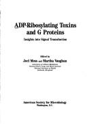
Adp-ribosylation. --- Bacterial toxins --- Industrial microorganisms --- Bacterial toxins. --- Guanine nucleotide regulatory protein. --- Poly adp ribose polymerase. --- Signal transduction. --- Adenosinedifosfaat. --- Biologische membranen. --- Stoftransport. --- Bacterial Toxins --- ADP Ribose Transferases --- Receptor-CD3 Complex, Antigen, T-Cell --- Receptor Cross-Talk --- Feedback, Physiological --- Gasotransmitters --- Toxins, Bacterial --- GTP-Binding Proteins --- Signal Transduction --- Receptor Mediated Signal Transduction --- Signal Transduction Pathways --- Signal Transduction Systems --- Receptor-Mediated Signal Transduction --- Signal Pathways --- Pathway, Signal --- Pathway, Signal Transduction --- Pathways, Signal --- Pathways, Signal Transduction --- Receptor-Mediated Signal Transductions --- Signal Pathway --- Signal Transduction Pathway --- Signal Transduction System --- Signal Transduction, Receptor-Mediated --- Signal Transductions --- Signal Transductions, Receptor-Mediated --- System, Signal Transduction --- Systems, Signal Transduction --- Transduction, Signal --- Transductions, Signal --- Cell Communication --- ADP Ribose Transferase --- ADPRT --- ADPRTs --- ART Transferase --- ART Transferases --- ARTase --- ARTases --- Mono ADP-ribose Transferases --- Mono ADPribose Transferase --- Mono ADPribose Transferases --- Mono(ADP-Ribose) Transferase --- Mono(ADP-Ribosyl)transferase --- Mono(ADPribosyl)transferase --- Mono-ADP-Ribosyltransferase --- MonoADPribosyltransferase --- NAD ADP-Ribosyltransferase --- NAD(+)-L-arginine ADP-D-ribosyltransferase --- NAD-Agmatine ADP-Ribosyltransferase --- NAD-Arginine ADP-Ribosyltransferase --- NADP-ADPRTase --- NADP-Arginine ADP-Ribosyltransferase --- ADP-Ribosyltransferase --- Mono(ADP-Ribose) Transferases --- NAD(P)(+)-Arginine ADP-Ribosyltransferase --- NAD+ ADP-Ribosyltransferase --- ADP Ribosyltransferase --- ADP-Ribosyltransferase, NAD --- ADP-Ribosyltransferase, NAD+ --- ADP-Ribosyltransferase, NAD-Agmatine --- ADP-Ribosyltransferase, NAD-Arginine --- ADP-Ribosyltransferase, NADP-Arginine --- ADP-ribose Transferases, Mono --- ADPribose Transferase, Mono --- ADPribose Transferases, Mono --- Mono ADP Ribosyltransferase --- Mono ADP ribose Transferases --- NAD ADP Ribosyltransferase --- NAD Agmatine ADP Ribosyltransferase --- NAD Arginine ADP Ribosyltransferase --- NAD+ ADP Ribosyltransferase --- NADP ADPRTase --- NADP Arginine ADP Ribosyltransferase --- Ribose Transferase, ADP --- Ribose Transferases, ADP --- Transferase, ADP Ribose --- Transferase, ART --- Transferase, Mono ADPribose --- Transferases, ADP Ribose --- Transferases, ART --- Transferases, Mono ADP-ribose --- Transferases, Mono ADPribose --- ADP-Ribosylation --- physiology --- ADP Ribose Transferases. --- Cellular signal transduction. --- G proteins --- Molecular microbiology --- Physiological effect. --- Genetics --- Congresses. --- Cell Signaling --- Bacterial Toxin --- Toxin, Bacterial --- Fungi. --- Antibiotics --- Lactones --- Streptomyces --- Yeasts
Book
ISBN: 9781627036368 1627036369 1627036377 Year: 2013 Publisher: Totowa, NJ : Humana Press : Imprint: Humana,
Abstract | Keywords | Export | Availability | Bookmark
 Loading...
Choose an application
- Reference Manager
- EndNote
- RefWorks (Direct export to RefWorks)
Featuring a diverse array of model organisms and scientific techniques, Sirtuins: Methods and Protocols collects detailed contributions from experts in the field addressing this vital family of genes. Opening with methods to generate sirtuin biology tools, the book continues by covering methods to identify sirtuin substrates, to measure sirtuin activity, and to study sirtuin biology. Written in the highly successful Methods in Molecular Biology series format, chapters include introductions to their respective topics, lists of the necessary materials and reagents, step-by-step, readily reproducible laboratory protocols, and tips on troubleshooting and avoiding known pitfalls. Comprehensive and easy to use, Sirtuins: Methods and Protocols presents detailed protocols for sirtuin research that can be followed directly or modified to investigate new areas of sirtuin biology.
Sirtuins. --- Proteins. --- Proteins --- Group III Histone Deacetylases --- Intracellular Signaling Peptides and Proteins --- ADP Ribose Transferases --- Histone Deacetylases --- Pentosyltransferases --- Amino Acids, Peptides, and Proteins --- Glycosyltransferases --- Amidohydrolases --- Transferases --- Chemicals and Drugs --- Hydrolases --- Enzymes --- Enzymes and Coenzymes --- Sirtuins --- Human Anatomy & Physiology --- Health & Biological Sciences --- Animal Biochemistry --- Silent Mating Type Information Regulator 2-like Proteins --- Sir2-like Proteins --- Silent Mating Type Information Regulator 2 like Proteins --- Sir2 like Proteins --- Coenzymes and Enzymes --- Biocatalysts --- Transferase --- Glycoside Transferases --- Transferases, Glycoside --- Gene Products, Protein --- Gene Proteins --- Protein Gene Products --- Proteins, Gene --- Class I Histone Deacetylases --- Class II Histone Deacetylases --- HDAC Proteins --- Histone Deacetylase --- Histone Deacetylase Complexes --- Complexes, Histone Deacetylase --- Deacetylase Complexes, Histone --- Deacetylase, Histone --- Deacetylases, Histone --- ADP Ribose Transferase --- ADPRT --- ADPRTs --- ART Transferase --- ART Transferases --- ARTase --- ARTases --- Mono ADP-ribose Transferases --- Mono ADPribose Transferase --- Mono ADPribose Transferases --- Mono(ADP-Ribose) Transferase --- Mono(ADP-Ribosyl)transferase --- Mono(ADPribosyl)transferase --- Mono-ADP-Ribosyltransferase --- MonoADPribosyltransferase --- NAD ADP-Ribosyltransferase --- NAD(+)-L-arginine ADP-D-ribosyltransferase --- NAD-Agmatine ADP-Ribosyltransferase --- NAD-Arginine ADP-Ribosyltransferase --- NADP-ADPRTase --- NADP-Arginine ADP-Ribosyltransferase --- ADP-Ribosyltransferase --- Mono(ADP-Ribose) Transferases --- NAD(P)(+)-Arginine ADP-Ribosyltransferase --- NAD+ ADP-Ribosyltransferase --- ADP Ribosyltransferase --- ADP-Ribosyltransferase, NAD --- ADP-Ribosyltransferase, NAD+ --- ADP-Ribosyltransferase, NAD-Agmatine --- ADP-Ribosyltransferase, NAD-Arginine --- ADP-Ribosyltransferase, NADP-Arginine --- ADP-ribose Transferases, Mono --- ADPribose Transferase, Mono --- ADPribose Transferases, Mono --- Mono ADP Ribosyltransferase --- Mono ADP ribose Transferases --- NAD ADP Ribosyltransferase --- NAD Agmatine ADP Ribosyltransferase --- NAD Arginine ADP Ribosyltransferase --- NAD+ ADP Ribosyltransferase --- NADP ADPRTase --- NADP Arginine ADP Ribosyltransferase --- Ribose Transferase, ADP --- Ribose Transferases, ADP --- Transferase, ADP Ribose --- Transferase, ART --- Transferase, Mono ADPribose --- Transferases, ADP Ribose --- Transferases, ART --- Transferases, Mono ADP-ribose --- Transferases, Mono ADPribose --- Intracellular Signaling Peptides --- Intracellular Signaling Proteins --- Peptides, Intracellular Signaling --- Proteins, Intracellular Signaling --- Signaling Peptides, Intracellular --- Signaling Proteins, Intracellular --- Proteids --- Amidases --- NAD-Dependent Histone Deacetylases --- Sir2-like Deacetylases --- Sirtuin Histone Deacetylases --- Deacetylases, NAD-Dependent Histone --- Deacetylases, Sir2-like --- Histone Deacetylases, NAD-Dependent --- NAD Dependent Histone Deacetylases --- Sir2 like Deacetylases --- Life sciences. --- Proteomics. --- Animal genetics. --- Life Sciences. --- Animal Genetics and Genomics. --- Genetics --- Molecular biology --- Biosciences --- Sciences, Life --- Science

ISBN: 9781588294999 1588294994 9781597450249 1617376027 9786610358526 1280358521 1597450243 Year: 2006 Publisher: Totowa, N.J. : Humana Press,
Abstract | Keywords | Export | Availability | Bookmark
 Loading...
Choose an application
- Reference Manager
- EndNote
- RefWorks (Direct export to RefWorks)
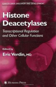
The recent discoveries that established histone acetylation as a key regulatory mechanism for gene expression triggered a wave of interest in histone posttranslational modifications and led to the development of novel anticancer agents now in clinical trials. In Histone Deacetylases: Transcriptional Regulation and Other Cellular Functions, a panel of leading investigators summarizes and synthesizes the new discoveries in this rapidly evolving field. The authors describe what has been learned about these proteins, including the identification of the enzymes, the elucidation of the enzymatic mechanisms of action, and the identification of their substrates and their partners. They also review the structures that have been solved for a number of enzymes-both alone and in complex with small-molecule inhibitors-and the biological roles of the several histone deacetylase (HDAC) genes that have been knocked out in mice. Authoritative and state-of-the-art, Histone Deacetylases: Transcriptional Regulation and Other Cellular Functions constitutes a first landmark of what has been accomplished so far and sets a clear agenda for the full definition of HDAC roles in biology and disease in the years to come.
Histone Deacetylases --- Cell Cycle --- Enzyme Repression --- Neoplasms --- Sirtuins --- Histone deacetylase. --- Cell cycle. --- Enzymes. --- Cancer --- Histone désacétylase --- Cycle cellulaire --- Enzymes --- physiology. --- drug effects. --- antagonists & inhibitors. --- drug therapy. --- Chemotherapy. --- Chimiothérapie --- Cancer -- Chemotherapy. --- Histone deacetylase --- Cell cycle --- Cell Physiological Processes --- Gene Expression Regulation, Enzymologic --- Enzyme Inhibitors --- ADP Ribose Transferases --- Amidohydrolases --- Group III Histone Deacetylases --- Biological Science Disciplines --- Intracellular Signaling Peptides and Proteins --- Diseases --- Therapeutics --- Analytical, Diagnostic and Therapeutic Techniques and Equipment --- Natural Science Disciplines --- Molecular Mechanisms of Pharmacological Action --- Pentosyltransferases --- Proteins --- Gene Expression Regulation --- Cell Physiological Phenomena --- Hydrolases --- Disciplines and Occupations --- Glycosyltransferases --- Amino Acids, Peptides, and Proteins --- Pharmacologic Actions --- Phenomena and Processes --- Genetic Processes --- Transferases --- Chemical Actions and Uses --- Enzymes and Coenzymes --- Genetic Phenomena --- Chemicals and Drugs --- Drug Therapy --- Histone Deacetylase Inhibitors --- Physiology --- Human Anatomy & Physiology --- Medicine --- Health & Biological Sciences --- Animal Biochemistry --- Oncology --- Chemotherapy --- Biocatalysts --- Ferments --- Soluble ferments --- Mitotic cycle --- Nuclear cycle (Cytology) --- Medicine. --- Cancer research. --- Biomedicine. --- Cancer Research. --- Antineoplastic agents --- Catalysts --- Enzymology --- Biological rhythms --- Amidases --- Treatment --- Oncology. --- Tumors --- Cancer research
Book
ISBN: 3642100112 3540884513 9786611986728 1281986720 3540884521 Year: 2009 Publisher: New York : Springer,
Abstract | Keywords | Export | Availability | Bookmark
 Loading...
Choose an application
- Reference Manager
- EndNote
- RefWorks (Direct export to RefWorks)
Vibrio cholerae, the causative organism of the disease Cholera, colonizes the small intestine and produces several different toxins among which the enterotoxin, or more widely known as cholera toxin (CT), happens to be the major virulence determinant that is responsible for the diarrheal syndrome. This book provides for the first time comprehensive and up-to-date information about all the toxins of Vibrio cholerae, their physical and chemical structures, their biosynthesis and its genetic regulation, their physiology, the molecular biology of their interactions with the host as well as their role in the development of an appropriate and effective cholera vaccine. It also offers relevant and necessary background information on the basic biology of the Vibrio cholerae cell and cholera bacteriophages.
Cholera toxin. --- Cholera toxin --- Bacterial Toxins --- ADP Ribose Transferases --- Enterotoxins --- Toxins, Biological --- Pentosyltransferases --- Biological Factors --- Glycosyltransferases --- Chemicals and Drugs --- Transferases --- Enzymes --- Enzymes and Coenzymes --- Cholera Toxin --- Biology --- Medicine --- Health & Biological Sciences --- Microbiology & Immunology --- Infectious Diseases --- Enterotoxins. --- Enterotoxin --- Cholera enterotoxin --- Cholera exotoxin --- Choleragen --- Choleragenoid --- CT (Cholera toxin) --- CTX (Cholera toxin) --- Procholeragenoid --- Vibrio cholerae enterotoxin --- Medicine. --- Medical microbiology. --- Pharmacology. --- Food --- Biochemistry. --- Cell biology. --- Microbiology. --- Biomedicine. --- Medical Microbiology. --- Food Science. --- Biochemistry, general. --- Cell Biology. --- Pharmacology/Toxicology. --- Biotechnology. --- Bacterial toxins --- NAD-ADP-ribosyltransferase --- Food science. --- Cytology. --- Toxicology. --- Chemicals --- Pharmacology --- Poisoning --- Poisons --- Cell biology --- Cellular biology --- Cells --- Cytologists --- Biological chemistry --- Chemical composition of organisms --- Organisms --- Physiological chemistry --- Chemistry --- Medical sciences --- Science --- Microbial biology --- Microorganisms --- Toxicology --- Composition --- Food—Biotechnology. --- Drug effects --- Medical pharmacology --- Chemotherapy --- Drugs --- Pharmacy --- Physiological effect
| Listing 1 - 6 of 6 |
Sort by
|

 Search
 Feedback
 About UniCat
  Help
Help News
News