| Listing 1 - 10 of 26 | << page >> |
Sort by
|
Book
ISBN: 2354030223 9782354030223 Year: 2009 Publisher: Paris: Med'Com,
Abstract | Keywords | Export | Availability | Bookmark
 Loading...
Choose an application
- Reference Manager
- EndNote
- RefWorks (Direct export to RefWorks)
Book
Year: 1991 Publisher: Chicago (Ill.) : University of Chicago press,
Abstract | Keywords | Export | Availability | Bookmark
 Loading...
Choose an application
- Reference Manager
- EndNote
- RefWorks (Direct export to RefWorks)

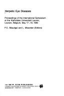
ISBN: 9061938015 9400986734 9400986718 Year: 1981 Publisher: The Hague Junk
Abstract | Keywords | Export | Availability | Bookmark
 Loading...
Choose an application
- Reference Manager
- EndNote
- RefWorks (Direct export to RefWorks)
Keratitis --- Academic collection --- Cornea --- Eye --- Inflammation --- Diseases --- Keratitis.

ISBN: 906193527X 9401089353 9400955189 Year: 1985 Volume: vol 44 Publisher: Dordrecht Boston Lancaster Junk
Abstract | Keywords | Export | Availability | Bookmark
 Loading...
Choose an application
- Reference Manager
- EndNote
- RefWorks (Direct export to RefWorks)
Keratitis, Dendritic. --- Simplexvirus --- Herpes simplex virus --- -Keratitis --- -Cornea --- Cornea --- Eye --- Herpesvirus hominis --- HSV (Virus) --- Herpesviruses --- Furrow Keratitis --- Keratitis, Furrow --- Dendritic Keratitides --- Dendritic Keratitis --- Furrow Keratitides --- Keratitides, Dendritic --- Keratitides, Furrow --- pathogenicity. --- Congresses --- Inflammation --- Diseases --- -pathogenicity. --- -Furrow Keratitis --- Keratitis --- Keratitis, Dendritic --- pathogenicity --- Hoornvliesontsteking. (Congres) --- Herpes simplex (Infection à). (Congrès) --- Kératite. (Congrès) --- Herpes simplex-infecties. (Congres) --- Herpes zoster - ophtalmicus
Book
ISBN: 9811052123 9811052115 Year: 2018 Publisher: Singapore : Springer Singapore : Imprint: Springer,
Abstract | Keywords | Export | Availability | Bookmark
 Loading...
Choose an application
- Reference Manager
- EndNote
- RefWorks (Direct export to RefWorks)
This book provides the concise descriptions of the basic and clinical knowledge about Acanthamoba keratitis, including characteristics of pathogen, risk factors, clinical manifestations, diagnosis and treatment with abundant figure illustrations and typical cases to ophthalmological practitioners and researchers. Acanthamoeba pathogen is widely distributed in the nature. However, Acanthamoeba keratitis is generally considered as a type of sight-threatening keratitis that is difficult to treat. At early stage Acanthamoeba keratitis usually shows atypical clinical manifestations, that are often misdiagnosed as viral or bacterial keratitis. Moreover, there are not approved topical anti-amoebicdrugs available up to now. We hope ophthalmological practitioners can obtain a comprehensive understanding of this infection through this book. Xuguang Sun is a professor of Ophthalmology at Beijing Institute of Ophthalmology, Beijing Tongren Eye Center, Beijing Tongren Hospital, Capital Medical University, Beijing, China.
Keratitis. --- Acanthamoeba. --- Medicine. --- Ophthalmology. --- Medicine & Public Health. --- Acanthamoebidae --- Cornea --- Eye --- Inflammation --- Diseases --- Medicine

ISBN: 0323037372 Year: 2006 Publisher: Philadelphia, PA : Elsevier Mosby,
Abstract | Keywords | Export | Availability | Bookmark
 Loading...
Choose an application
- Reference Manager
- EndNote
- RefWorks (Direct export to RefWorks)
Conjunctivitis. --- Eye --- Keratitis. --- Uveitis. --- Eye Diseases --- Inflammation --- Diseases. --- Inflammation. --- Immunology. --- Therapy.
Dissertation
Abstract | Keywords | Export | Availability | Bookmark
 Loading...
Choose an application
- Reference Manager
- EndNote
- RefWorks (Direct export to RefWorks)
Keratitis --- Corneal Stroma --- Cornea --- Aldehyde Dehydrogenase --- Antigen-Antibody Complex --- immunology --- chemistry --- metabolism
Dissertation
Year: 1986 Publisher: Amsterdam Rodopi
Abstract | Keywords | Export | Availability | Bookmark
 Loading...
Choose an application
- Reference Manager
- EndNote
- RefWorks (Direct export to RefWorks)
Keratitis, Dendritic --- Eyelid Diseases --- Simplexvirus --- Keratoconjunctivitis --- microbiology --- drug therapy --- isolation & purification --- etiology
Book
ISBN: 9783642144875 9783642144868 Year: 2011 Publisher: Berlin Heidelberg Springer Berlin Heidelberg Imprint Springer
Abstract | Keywords | Export | Availability | Bookmark
 Loading...
Choose an application
- Reference Manager
- EndNote
- RefWorks (Direct export to RefWorks)
Herpes zoster ophthalmicus (HZO) is a common disease in the elderly and the immunosuppressed, with potentially devastating sequelae. Diagnosis of HZO is clinical but almost all its manifestations are non-specific and often indistinguishable from those due to other causes in general and herpes simplex virus in particular. The exception is varicella-zoster virus epithelial keratitis, which is frequently the only indicator of the true nature of the disease. This book is unique in presenting high-magnification images, obtained by non-contact in vivo photomicrography, that capture the distinctive features of varicella-zoster virus epithelial keratitis in HZO. Both the morphology and the dynamics of the corneal epithelial lesions are splendidly documented, including in patients with HZO sine herpete and recurrent disease. Three rare cases of ocular surface involvement in acute HZO are included, and the final chapter offers an illuminating comparison of varicella-zoster virus epithelial keratitis in HZO and the lesions of herpes simplex virus. This book will serve as an indispensable aid in the prompt diagnosis of HZO.
Infectious diseases. Communicable diseases --- Ophthalmology --- besmettelijke ziekten --- oftalmologie --- Herpes Zoster Ophthalmicus --- Herpesvirus 3, Human. --- Keratitis, Herpetic --- Varicella-zoster virus --- Keratitis --- Ophthalmic zoster --- Virus varicello-zonateux --- Kératite --- Zona ophtalmique --- physiopathology. --- EPUB-LIV-FT LIVMEDEC SPRINGER-B
Book
ISBN: 9783319065458 3319065440 9783319065441 3319065459 Year: 2015 Publisher: Cham : Springer International Publishing : Imprint: Springer,
Abstract | Keywords | Export | Availability | Bookmark
 Loading...
Choose an application
- Reference Manager
- EndNote
- RefWorks (Direct export to RefWorks)
This book on the morphology of corneal surface changes in recurrent erosion syndrome and epithelial edema presents high-magnification images captured in vivo by the method of non-contact photomicrography. Part I of the book, on recurrent erosion syndrome, displays images covering a broad spectrum of epithelial changes, including manifestations of the ongoing underlying pathological process and epithelial activity aimed at elimination of abnormal elements and repair. The dynamics of the interplay between these opposing forces are captured in sequential photographs that are invaluable for interpretation. Case reports illustrate typical features of the disease and document the variability of symptoms and findings in the same individual over time. Also included are images of the appearance and dynamics of corneal stromal infiltrates, a rare but potentially sight-threatening complication. Part II of the book demonstrates typical features of corneal epithelial edema and also covers the occasional contemporaneous occurrence, and dynamics, of various phenomena indistinguishable from those commonly seen in recurrent erosion syndrome. Again, informative case reports are included. The in vivo images displayed in this book, obtained at a higher magnification than that used in standard photography, reveal additional details of epithelial changes. The presented morphology will facilitate understanding of clinical appearances and assist in differential diagnosis.
Medicine & Public Health. --- Ophthalmology. --- Medicine. --- Médecine --- Ophtalmologie --- Medicine --- Health & Biological Sciences --- Ophthalmology & Optometry --- Cornea --- Keratitis. --- Eye --- Diseases. --- Keratomalacia --- Diseases and defects --- Inflammation --- Ophthalmology --- Diseases
| Listing 1 - 10 of 26 | << page >> |
Sort by
|

 Search
 Feedback
 About UniCat
  Help
Help News
News