| Listing 1 - 3 of 3 |
Sort by
|

ISBN: 1280462531 9786610462537 3540281665 3540281622 Year: 2006 Publisher: Berlin : Springer,
Abstract | Keywords | Export | Availability | Bookmark
 Loading...
Choose an application
- Reference Manager
- EndNote
- RefWorks (Direct export to RefWorks)
Pain is an unpleasant but very important biological signal for danger. Nociception is necessary for survival and maintaining the integrity of the organism in a potentially hostile environment. Pain is both a sensory experience and a perceptual metaphor for damage and it is activated by noxious stimuli that act on a complex pain sensory apparatus. However, chronic pain having no more a protective role can become a ruining disease itself, termed "neuropathic pain".
Pain. --- Nociceptors. --- Neuralgia. --- Pain receptors --- Sensory receptors --- Aches --- Emotions --- Pleasure --- Senses and sensation --- Symptoms --- Analgesia --- Suffering --- Nerves, Peripheral --- Pain --- Diseases --- Neurosciences. --- Neural sciences --- Neurological sciences --- Neuroscience --- Medical sciences --- Nervous system
Book
ISBN: 1281862479 9786611862473 354079462X 3540794611 Year: 2008 Publisher: Berlin : Springer,
Abstract | Keywords | Export | Availability | Bookmark
 Loading...
Choose an application
- Reference Manager
- EndNote
- RefWorks (Direct export to RefWorks)
This monograph gives an overview of the STN. It treats the position of the STN in hemiballism, based on older and recent data. The cytology encompasses the neuronal types present in the STN in nearly all studied species and focuses on interneurons and the extent of their dendrites. Ultrastructural features are described for cat and baboon (F1, F2, Sr, LR1, LR2 boutons and d.c.v. terminals, together with vesicle containing dendrites), the cytochemistry is focused on receptors (dopamine, cannabinoid, opioid, glutamate, GABA, serotonin, and cholinergic-, purinergic ones) and calcium binding proteins and calcium channels. The development of the subthalamic nucleus from the subthalamic cell cord is given together with its developing connections. The topography of rat, cat, baboon and man is worked out as to cytology, sagittal borders, surrounding nuclei and tracts, and aging of the human STN. The connections of the STN are extensively elaborated on: cortical-, subthalamo-cortical-, pallidosubthalamic-, pedunculopontine-, raphe-, thalamic-, central grey-, and nigral connections. Emphasis is put on human connections. Recent nigro-subthalamic studies showed a contralateral projection. The role of the STN in the basal ganglia circuitry is described as to the direct, indirect and hyperdirect pathway. The change the STN undergoes in Parkinson’s disease in neuronal firing rate and firing pattern is demonstrated together with the possible mechanisms of deep brain stimulation. The results of in vitro measurements on dissociated cultured subthalamic neurons are presented. The preliminary effects of application of acetylcholine and high frequency stimulation are described. This part is preceded with studies concerning spontaneous activity, depolarizing and hyperpolarizing inputs, synaptic inputs, high frequency stimulation, and burst activity of STN cells. The last extensive part concerns STN cell models and simulation of neuronal networks. Single cell models (model of Otsuka and Terman/Rubin) are compared and the multi-compartment model of Gillies and Willshaw is explored. The globus pallidus externus-STN network as proposed by Terman is briefly described. The monograph finishes with a series of interpretations of the results.
Subthalamus. --- Subthalamus --- Basal ganglia. --- Neural networks (Neurobiology) --- Cytology. --- Biological neural networks --- Nets, Neural (Neurobiology) --- Networks, Neural (Neurobiology) --- Neural nets (Neurobiology) --- Cognitive neuroscience --- Neurobiology --- Neural circuitry --- Ganglia, Basal --- Efferent pathways --- Extrapyramidal tracts --- Telencephalon --- Corpus subthalamicum --- Brain --- Diencephalon --- Neurosciences. --- Neural sciences --- Neurological sciences --- Neuroscience --- Medical sciences --- Nervous system

ISBN: 3540280448 9786610462520 1280462523 3540280456 Year: 2006 Publisher: Berlin ; New York : Springer,
Abstract | Keywords | Export | Availability | Bookmark
 Loading...
Choose an application
- Reference Manager
- EndNote
- RefWorks (Direct export to RefWorks)
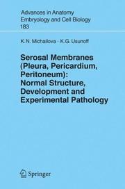
in the human visceral pleura is the sole reliable criterion for the statement that it belongs to the ‘thick type’, while all observed animals have a ‘thin’ type VP. The mesothelium and underlying structures of the SM represent a highly permeable bidirectional membrane with signi?cant differences in the organ and region tra- port as a local phenomenon after horseradish peroxidase application. Stomata are constant features seen by SEM, but are occasional ?ndings observed by TEM over both sides of the diaphragm, lower intercostal spaces, anterior abdominal wall and greater omentum in untreated animals. Our data extend the location of stomata over the liver and broad ligament of the uterus. We strictly de?ned and nominated the main structures of the lymphatic regions as lymphatic units, stomata, and LL. Several different types of vascularization of omental and extraomental (medias- nal pleura and lesser pelvis) MS are observed after India ink application. Human and animal differences in their location, mesothelial covering, the vessel (blood and lymphatic) supply, free and connective tissue cells and their arrangement are discussed. The mesothelium and the BL changes start early in the gestation and continue throughout the postnatal period. Both cell types (?at and cubic) are already evident through prenatal life.
Anatomy. --- Embryology. --- Membranes (Biology). --- Serous fluids. --- Membranes --- Tissues --- Anatomy --- Serous Membrane --- Human Anatomy & Physiology --- Biology --- Zoology --- Health & Biological Sciences --- Cytology --- Animal Anatomy & Embryology --- Physiology --- Membranes (Biology) --- Serosal fluids --- Biological membranes --- Biomembranes --- Medicine. --- Human physiology. --- Biomedicine. --- Human Physiology. --- Lymph --- Biological interfaces --- Protoplasm --- Human biology --- Medical sciences --- Human body
| Listing 1 - 3 of 3 |
Sort by
|

 Search
 Feedback
 About UniCat
  Help
Help News
News