| Listing 1 - 5 of 5 |
Sort by
|
Book
ISBN: 9783319096599 3319096583 9783319096582 3319096591 Year: 2015 Publisher: Cham : Springer International Publishing : Imprint: Springer,
Abstract | Keywords | Export | Availability | Bookmark
 Loading...
Choose an application
- Reference Manager
- EndNote
- RefWorks (Direct export to RefWorks)
Based on the experience of two Italian referral centers, the book depicts the characteristic findings obtained when using MR imaging to study the male and female pelvis including the obstetric applications. Each chapter provides a comprehensive account of the use of the imaging technique of examination, including the most recent advances in MR imaging, the anatomy and MR possibilities in the identification, characterization and staging of the different pelvic diseases highlighting its diagnostic possibilities. The advances in fetal MRI, representing the cutting edge of pelvic MR imaging, will also be depicted. The text is complemented by numerous illustrations, as well as clinical cases that make this a very practice-oriented work, presenting the role of diagnostic imaging in every-day clinical activity. The volume will prove an invaluable guide for both residents and professionals with core interest in gynecology, obstetrics and urology.
Medicine & Public Health. --- Imaging / Radiology. --- Gynecology. --- Obstetrics/Perinatology. --- Urology/Andrology. --- Medicine. --- Obstetrics. --- Radiology, Medical. --- Urology. --- Médecine --- Gynécologie --- Obstétrique --- Urologie --- Magnetic resonance imaging. --- Medicine --- Health & Biological Sciences --- Radiology, MRI, Ultrasonography & Medical Physics --- Pelvis --- Magnetic resonance imaging --- Pelvic cavity --- Pelvic region --- Radiology. --- Anatomy --- Obstetrics/Perinatology/Midwifery. --- Genitourinary organs --- Maternal-fetal medicine --- Gynaecology --- Generative organs, Female --- Clinical radiology --- Radiology, Medical --- Radiology (Medicine) --- Medical physics --- Diseases --- Gynecology . --- Radiological physics --- Physics --- Radiation
Book
ISBN: 8847028434 8847028442 9788847028432 Year: 2013 Publisher: Milano : Springer Milan : Imprint: Springer,
Abstract | Keywords | Export | Availability | Bookmark
 Loading...
Choose an application
- Reference Manager
- EndNote
- RefWorks (Direct export to RefWorks)
This book, based on the experience of a single large referral center, presents the characteristic findings obtained when using MR imaging and MR cholangiopancreatography (MRCP) to image the biliary tree and pancreatic ducts in a variety of disease settings. An introductory chapter is devoted to technical considerations, anatomy, and developmental anomalies. Subsequent chapters then present in detail the MR imaging and MRCP findings observed in choledocholithiasis, inflammatory and neoplastic disorders of the bile ducts, acute and chronic pancreatitis (according to etiology), and different pancreatic neoplasms. Dynamic MRCP with secretin stimulation is also illustrated, documenting both normal and abnormal responses of the pancreatic duct system to secretin. Readers will find this book to be an excellent aid to the interpretation of MR imaging and MRCP findings in patients with biliary and pancreatic disease.
Biliary tract -- Magnetic resonance imaging. --- Endoscopic retrograde cholangiopancreatography. --- Biliary tract --- Magnetic Resonance Imaging --- Diagnostic Techniques, Digestive System --- Digestive System --- Pancreas --- Anatomy --- Tomography --- Diagnostic Techniques and Procedures --- Diagnostic Imaging --- Diagnosis --- Analytical, Diagnostic and Therapeutic Techniques and Equipment --- Biliary Tract --- Cholangiopancreatography, Magnetic Resonance --- Pancreatic Ducts --- Medicine --- Health & Biological Sciences --- Gastroenterology --- Radiology, MRI, Ultrasonography & Medical Physics --- Magnetic resonance imaging --- Magnetic resonance imaging. --- Cholangiopancreatography, Endoscopic retrograde --- ERCP (Radiography) --- Biliary tree --- Medicine. --- Radiology. --- Gastroenterology. --- Hepatology. --- Medicine & Public Health. --- Diagnostic Radiology. --- Endoscopy --- Pancreatic duct --- Digestive organs --- Bile --- Radiography --- Radiology, Medical. --- Clinical medicine. --- Medicine, Clinical --- Internal medicine --- Clinical radiology --- Radiology, Medical --- Radiology (Medicine) --- Medical physics --- Diseases --- Gastroenterology . --- Radiological physics --- Physics --- Radiation --- Biliary tract. --- Pancreatic duct. --- Voies biliaires --- Voies biliaires. --- Canal de Wirsung. --- Imagerie par résonance magnétique.
Digital
ISBN: 9783319096599 9783319096605 9783319096582 9783319355597 Year: 2015 Publisher: Cham Springer International Publishing
Abstract | Keywords | Export | Availability | Bookmark
 Loading...
Choose an application
- Reference Manager
- EndNote
- RefWorks (Direct export to RefWorks)
Based on the experience of two Italian referral centers, the book depicts the characteristic findings obtained when using MR imaging to study the male and female pelvis including the obstetric applications. Each chapter provides a comprehensive account of the use of the imaging technique of examination, including the most recent advances in MR imaging, the anatomy and MR possibilities in the identification, characterization and staging of the different pelvic diseases highlighting its diagnostic possibilities. The advances in fetal MRI, representing the cutting edge of pelvic MR imaging, will also be depicted. The text is complemented by numerous illustrations, as well as clinical cases that make this a very practice-oriented work, presenting the role of diagnostic imaging in every-day clinical activity. The volume will prove an invaluable guide for both residents and professionals with core interest in gynecology, obstetrics and urology.
Physical methods for diagnosis --- Urology. Andrology --- Gynaecology. Obstetrics --- vrouwen --- obstetrie --- urologie --- perinatale sterfte --- gynaecologie --- vroedkunde --- radiologie --- medische beeldvorming

ISBN: 9783540682516 9783540002819 Year: 2009 Publisher: Berlin Heidelberg Springer Berlin Heidelberg
Abstract | Keywords | Export | Availability | Bookmark
 Loading...
Choose an application
- Reference Manager
- EndNote
- RefWorks (Direct export to RefWorks)
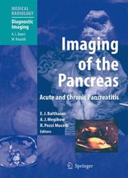
With the aid of numerous high-quality illustrations, this volume explains the strengths and limitations of the different techniques employed in the imaging of pancreatitis. Ultrasound, computed tomography, magnetic resonance imaging and interventional imaging are each considered separately in the settings of acute and chronic pancreatitis. A further section is devoted to imaging of the complications of these conditions. Throughout, care has been taken to ensure that the reader will achieve a sound understanding of how the imaging findings derive from the pathophysiology of the disease processes. The significance of the imaging findings for clinical and therapeutic decision making is clearly explained, and protocols are provided that will assist in obtaining the best possible images.
Medicine & Public Health. --- Imaging / Radiology. --- Diagnostic Radiology. --- Abdominal Surgery. --- Gastroenterology. --- Oncology. --- Medicine. --- Medical radiology --- Gastroenterology. --- Oncology. --- Abdomen --- Médecine --- Radiologie médicale --- Gastroentérologie --- Cancérologie --- Abdomen --- Surgery. --- Chirurgie
Multi
ISBN: 9788847017696 9788847017689 9788847017702 Year: 2010 Publisher: Milano Springer
Abstract | Keywords | Export | Availability | Bookmark
 Loading...
Choose an application
- Reference Manager
- EndNote
- RefWorks (Direct export to RefWorks)
La presente pubblicazione dedicata alla patologia non oncologica dell'Apparato Urogenitale nasce da un'idea del nucleo storico degli Uroradiologi italiani, in particolare del Prof. Antonio Rotondo, i quali si occupano da anni dell'argomento e che, sotto l'egida della Sezione di studio di radiologia urogenitale, hanno coinvolto autori, giovani e meno giovani, ma comunque rappresentativi degli addetti ai lavori, esperti di questa branca specialistica. Il testo intende fornire informazioni sullo stato dell'arte, che possano essere utili nella clinica, sia a chi pratica quotidianamente la disciplina sia a chi se ne occupa saltuariamente. L'opera è corredata da una ricca e significativa iconografia così come da schemi di semplificazione, che consentono di accedere rapidamente ai concetti fondamentali delle varie problematiche.
Physical methods for diagnosis --- Urology. Andrology --- urologie --- radiologie --- medische beeldvorming
| Listing 1 - 5 of 5 |
Sort by
|

 Search
 Feedback
 About UniCat
  Help
Help News
News