| Listing 1 - 5 of 5 |
Sort by
|

ISBN: 1281352195 9786611352196 354033677X 3540336753 Year: 2007 Publisher: Berlin, Heidelberg : Springer Berlin Heidelberg : Imprint: Springer,
Abstract | Keywords | Export | Availability | Bookmark
 Loading...
Choose an application
- Reference Manager
- EndNote
- RefWorks (Direct export to RefWorks)
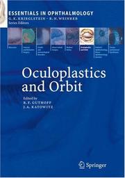
The series Essentials in Ophthalmology was initiated- to discuss clinically relevant and appropriate topics two years ago to expedite the timely transfer. Summaries of clinically relevant information of new information in vision science and have been provided throughout each chapter. evidence-based medicine into clinical practice. Each subspecialty area now has been covered We thought that this prospicient idea would be once, and the response to the first eight volumes moved and guided by a resolute commitment in the series has been enthusiastically positive. to excellence. It is reasonable to now update our With the start of the second cycle of subspecialty readers with what has been achieved. coverage, the dissemination of practical inform- a The immediate goal was to transfer information will be continued as we learn more about through a high quality quarterly publication the emerging advances in various ophthalmic in which ophthalmology would be represented by subspecialties that can be applied to obtain the eight subspecialties. In this regard, each issue has best possible care of our patients. Moreover, we had a subspecialty theme and has been overseen will continue to highlight clinically relevant - by two internationally recognized volume editors- formation and maintain our commitment to editors, who in turn have invited a bevy of experts excellence. G. K. Krieglstein R. N.
Ophthalmic plastic surgery --- Adnexa oculi --- Eye-sockets --- Technique. --- Diseases --- Surgery --- Accessory organs of the eye --- Anterior adnexa --- Ocular adnexa --- Organa oculi accessoria --- Eye --- Orbit (Eye) --- Orbital cavity (Eye) --- Facial bones --- Ocular plastic surgery --- Oculoplastic surgery --- Oculoplasty --- Ophthalmoplastic surgery --- Ophthalmoplasty --- Surgery, Plastic --- Ophthalmology. --- Surgery. --- Otorhinolaryngology. --- Endoscopic surgery. --- Plastic Surgery. --- Oral and Maxillofacial Surgery. --- Minimally Invasive Surgery. --- Endosurgery --- Minimal access surgery --- Minimally invasive surgery --- MIS (Minimally invasive surgery) --- Operative endoscopy --- Surgical endoscopy --- Endoscopy --- Microsurgery --- Surgery, Operative --- Ear, nose, and throat diseases --- ENT diseases --- Otorhinolaryngology --- Medicine --- Surgery, Primitive --- Plastic surgery. --- Oral surgery. --- Maxillofacial surgery. --- Minimally invasive surgery. --- Dental surgery --- Oral surgery --- Surgery, Dental --- Surgery, Oral --- Oral surgeons --- Aesthetic surgery --- Cosmetic surgery --- Plastic surgery --- Reconstructive surgery --- Surgery, Aesthetic --- Surgery, Cosmetic --- Surgery, Reconstructive --- Transplantation of organs, tissues, etc. --- Plastic surgeons

ISBN: 1280863927 9786610863921 354032707X 3540327053 Year: 2006 Publisher: Berlin Heidelberg : Springer-Verlag GmbH,
Abstract | Keywords | Export | Availability | Bookmark
 Loading...
Choose an application
- Reference Manager
- EndNote
- RefWorks (Direct export to RefWorks)
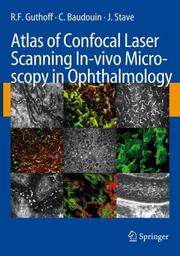
Confocal microscopy with laser scanning technology yields in-vivo images of ocular and ocular adnexal surfaces that are so brilliant that they rival histology in terms of quality.This unique atlas and textbook demonstrates normal in-vivo anatomy of the cornea, limbus and conjunctiva, quantifies various cellular structures using cell-density calculations and establishes correlations between novel optical sections of various diseases of the ocular surface and clinical findings. Furthermore, it supports the interpretation of novel high-magnification optical sections by comparing corneal and conjunctival imprint cytology with in-vivo images and describes early inflammatory changes in corneal grafts, as well as corneal conjunctivalisation in limbal stem cell deficiency, corneal dystrophies or infections, flap interface and margin characteristics after laser in-situ keratomileusis (LASIK). In addition, it instructs the reader about diagnostic and therapeutic follow-up strategies and provides a brief introduction to applications in other fields such as dentistry and ear, nose and throat surgery.
Confocal microscopy. --- Ophthalmology. --- Otolaryngology. --- Otorhinolaryngology. --- Dentistry. --- Dermatology. --- Biological and Medical Physics, Biophysics. --- Medicine --- Eye --- Skin --- Dental surgery --- Odontology --- Surgery, Dental --- Oral medicine --- Teeth --- Ear, nose, and throat diseases --- ENT diseases --- Otorhinolaryngology --- Diseases --- Biophysics. --- Biological physics. --- Biological physics --- Biology --- Medical sciences --- Physics
Digital
ISBN: 9783540327073 Year: 2006 Publisher: Berlin Heidelberg Springer-Verlag GmbH
Abstract | Keywords | Export | Availability | Bookmark
 Loading...
Choose an application
- Reference Manager
- EndNote
- RefWorks (Direct export to RefWorks)
General biophysics --- Otorhinolaryngology --- Stomatology --- Pathological dermatology --- Ophthalmology --- biofysica --- dermatologie --- otorinolaryngologie --- oftalmologie --- stomatologie
Book
ISBN: 9783540327073 Year: 2006 Publisher: Berlin Heidelberg Springer-Verlag GmbH.
Abstract | Keywords | Export | Availability | Bookmark
 Loading...
Choose an application
- Reference Manager
- EndNote
- RefWorks (Direct export to RefWorks)
Confocal microscopy with laser scanning technology yields in-vivo images of ocular and ocular adnexal surfaces that are so brilliant that they rival histology in terms of quality.This unique atlas and textbook demonstrates normal in-vivo anatomy of the cornea, limbus and conjunctiva, quantifies various cellular structures using cell-density calculations and establishes correlations between novel optical sections of various diseases of the ocular surface and clinical findings. Furthermore, it supports the interpretation of novel high-magnification optical sections by comparing corneal and conjunctival imprint cytology with in-vivo images and describes early inflammatory changes in corneal grafts, as well as corneal conjunctivalisation in limbal stem cell deficiency, corneal dystrophies or infections, flap interface and margin characteristics after laser in-situ keratomileusis (LASIK). In addition, it instructs the reader about diagnostic and therapeutic follow-up strategies and provides a brief introduction to applications in other fields such as dentistry and ear, nose and throat surgery.
General biophysics --- Otorhinolaryngology --- Stomatology --- Pathological dermatology --- Ophthalmology --- biofysica --- dermatologie --- otorinolaryngologie --- oftalmologie --- stomatologie
Book

ISBN: 9783110851854 Year: 2020 Publisher: Berlin Boston
Abstract | Keywords | Export | Availability | Bookmark
 Loading...
Choose an application
- Reference Manager
- EndNote
- RefWorks (Direct export to RefWorks)
| Listing 1 - 5 of 5 |
Sort by
|

 Search
 Feedback
 About UniCat
  Help
Help News
News