
 Search
 Feedback
 About UniCat
  Help
Help News
News
| Listing 1 - 4 of 4 |
Sort by
|
Book
ISBN: 3642178715 9786613369192 1283369192 3642178723 Year: 2011 Publisher: Berlin : Springer,
Abstract | Keywords | Export | Availability | Bookmark
 Loading...
Choose an application
- Reference Manager
- EndNote
- RefWorks (Direct export to RefWorks)
Imaging is increasingly used in patients suspected of having acute appendicitis in order to reduce the rates of perforation and negative appendectomy and to detect alternative diagnoses. As many of these patients are young, radiation dose is of particular concern. Numerous studies have therefore investigated reduction of the radiation dose delivered by CT and whether ultrasound may be a preferable option, depending on age and gender. It is also recognized that MRI can be invaluable in certain patients, such as pregnant women, though it is difficult to use in emergency settings. This book is a comprehensive account of imaging of acute appendicitis and other appendiceal diseases. Background information is first provided on clinical presentation, perforation and negative appendectomy rates, and treatment options. The role of each imaging modality is then considered separately in adults and children with suspected acute appendicitis. Many high-quality illustrations are included, and detailed information is provided on appropriate protocols and radiation saving. Further chapters addresses the spontaneously resolving and chronic appendicitis as well as other appendiceal lesions, and review the findings of evidence-based medicine and cost-effectiveness analyses. Emergency physicians, pediatricians, surgeons, and radiologists will all find this book to be an excellent source of information and guidance.
Appendicitis -- Imaging. --- Appendix (Anatomy) -- Imaging. --- Appendix (Anatomy) --- Appendicitis --- Diagnostic Techniques and Procedures --- Cecal Diseases --- Gastroenteritis --- Investigative Techniques --- Intestinal Diseases --- Gastrointestinal Diseases --- Analytical, Diagnostic and Therapeutic Techniques and Equipment --- Diagnosis --- Digestive System Diseases --- Diseases --- Methods --- Diagnostic Imaging --- Medicine --- Surgery & Anesthesiology --- Health & Biological Sciences --- Radiology, MRI, Ultrasonography & Medical Physics --- Surgery - General and By Type --- Imaging --- Ultrasonic imaging. --- Imaging. --- Perityphlitis --- Medicine. --- Emergency medicine. --- Radiology. --- Abdominal surgery. --- Medicine & Public Health. --- Imaging / Radiology. --- Diagnostic Radiology. --- Ultrasound. --- Emergency Medicine. --- Abdominal Surgery. --- Inflammation --- Radiology, Medical. --- Diagnosis, Ultrasonic. --- Abdomen --- Surgery. --- Abdominal surgery --- Laparotomy --- Medicine, Emergency --- Critical care medicine --- Disaster medicine --- Medical emergencies --- Diagnosis, Ultrasonic --- Diagnostic sonography --- Diagnostic ultrasonics --- Diagnostic ultrasonography --- Diagnostic ultrasound --- Medical diagnostic ultrasonic imaging --- Medical ultrasonography --- Ultrasonic diagnosis --- Ultrasonic diagnostic imaging --- Ultrasonic imaging --- Ultrasonic waves --- Diagnostic imaging --- Ultrasonics in medicine --- Clinical radiology --- Radiology, Medical --- Radiology (Medicine) --- Medical physics --- Diagnostic use --- Radiological physics --- Physics --- Radiation
ISBN: 1280462574 9786610462575 3540309039 3540213430 Year: 2006 Publisher: Berlin ; London : Springer,
Abstract | Keywords | Export | Availability | Bookmark
Loading...Choose an application
- Reference Manager
- EndNote
- RefWorks (Direct export to RefWorks)
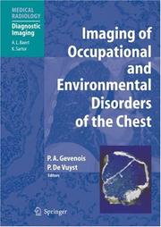
The spectrum of occupational and environmental diseases has changed markedly during the past decade. Pneumoconiosis is still a common cause of chronic lung disease but the concept has evolved to encompass reactions of the lung to chemicals and organic or inorganic dust. Furthermore, new industrial processes have led to the production and use of a wide range of chemicals, metals, and alloys, an increasing number of which, including man-made organic particles, are reported to cause interstitial lung disease in exposed workers. Thus, while the workforce in coal mining and asbestos handling has decreased, new groups of workers are at risk of exposure to large amounts of mineral and metallic dusts that may induce pneumoconiosis. This well-illustrated book, written by internationally acclaimed experts, provides an up-to-date and comprehensive approach to modern imaging of environmental and occupational diseases of the chest. The first part of the book addresses the basic knowledge required to understand imaging in this context, while the second focuses on the imaging results achieved in a variety of specific disorders. There is particular emphasis on the role of thin-section computed tomography since this technique facilitates the detection of early subclinical abnormalities.
Occupational diseases --- Environmentally induced diseases --- Chest --- Diagnosis. --- Imaging. --- Diseases --- Environmental aspects. --- Thorax, Human --- Thorax (Zoology) --- Viscera --- Clinical ecology --- Environmental illness --- Environmental health --- Medical geography --- Environmental aspects --- Causes and theories of causation --- Radiology, Medical. --- Pneumology. --- Medicine, Industrial. --- Imaging / Radiology. --- Diagnostic Radiology. --- Pneumology/Respiratory System. --- Occupational Medicine/Industrial Medicine. --- Industrial medicine --- Medicine, Occupational --- Occupational medicine --- Medicine --- Clinical radiology --- Radiology, Medical --- Radiology (Medicine) --- Medical physics --- Radiology. --- Respiratory organs—Diseases. --- Occupational medicine. --- Radiological physics --- Physics --- Radiation --- Respiratory organs --- Occupational health services. --- Occupational Health. --- Diseases. --- Employee health services --- Medical care --- Medicine, Industrial --- Respiratory diseases
Book
ISBN: 9783642245350 9786613939296 3642245358 364224534X 1283626845 Year: 2012 Publisher: Heidelberg ; New York : Springer,
Abstract | Keywords | Export | Availability | Bookmark
Loading...Choose an application
- Reference Manager
- EndNote
- RefWorks (Direct export to RefWorks)
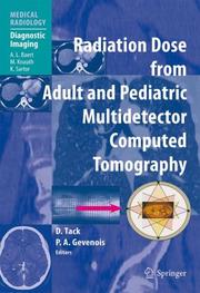
Computed tomography (CT) is a powerful technique providing precise and confident diagnoses. Close to 65 million CT examinations are performed each year in the United States alone, an astonishing number achieved with an annual double digit growth rate over the past 30 years. The burgeoning use of CT has resulted in an exponential increase in collective radiation dose to the population. Despite investigations supporting the use of lower radiation doses, surveys highlight the lack of proper understanding of CT parameters that affect radiation dose. Dynamic advances in CT technology also make it important to explain the latest dose-saving strategies in an easy-to-comprehend manner relevant to routine clinical practice. This book aims to review all aspects of the radiation dose from CT and to provide simple rules and tricks for radiologists and radiographers that will assist in the appropriate use of CT technique. The second edition includes a number of new chapters on the most up-to-date strategies and technologies for radiation dose reduction while updating the outstanding contents of the first edition. Vendor perspectives are included, and an online image gallery will also be available to readers.
Radiotherapy. Isotope therapy --- Paediatrics --- Physical methods for diagnosis --- Neuropathology --- Human medicine --- neurologie --- farmacologie --- geneeskunde --- pediatrie --- radiologie --- medische beeldvorming --- Radiology, Medical. --- Pediatrics. --- Internal medicine. --- Medicine, Internal --- Medicine --- Pediatric medicine --- Children --- Clinical radiology --- Radiology, Medical --- Radiology (Medicine) --- Medical physics --- Diseases --- Health and hygiene --- Radiology. --- Neuroradiology. --- Imaging / Radiology. --- Diagnostic Radiology. --- Internal Medicine. --- Neuroradiography --- Neuroradiology --- Nervous system --- Radiological physics --- Physics --- Radiation --- Radiography. --- Tomography --- Safety measures.
ISBN: 1281167622 9786611167622 3540685758 3540288880 3642066941 Year: 2007 Publisher: Berlin : Springer,
Abstract | Keywords | Export | Availability | Bookmark
Loading...Choose an application
- Reference Manager
- EndNote
- RefWorks (Direct export to RefWorks)
The number of computed tomography (CT) examinations has increased continuously since 1980 for a variety of reasons, including new indications and growth in the number of CT units. Beyond its use in adults with suspected malignancies and other conditions, CT is now used in young patients, including children, who are suffering from benign diseases and have a long life expectancy. The consequence of this trend is that CT, and in particular multidetector row CT (MDCT), is now responsible for about two-thirds of the total radiation dose delivered to the population for diagnostic purposes. Because of the associated radiation dose, CT may induce cancers, and the risk of death has been estimated at up to one per 1,000 CT examinations. Even if the balance between the risks related to ionizing radiation and the benefits from an accurate diagnosis provided by CT scanning has always been considered highly positive, it is now mandatory to reduce and/or to optimize the radiation dose. Referring clinicians, radiologists, and technologists should therefore all understand the mechanisms that determine the radiation risk, but it has been shown that only a small proportion of these professionals are aware of both the radiation risks and their underlying mechanisms. This book is designed to rectify this situation. The first part of the book provides a comprehensive approach to all the factors that influence the radiation dose and subsequently the risk induced by using MDCT in children and adult patients. In the second part, guidelines are proposed for optimization of the radiation dose in order to obtain an image quality sufficient for appropriate diagnostic performance while restricting the dose delivered. The authors are experts of international standing, selected for their acknowledged scientific contributions. This book will appeal to both general and specialized radiologists, including pediatric radiologists, CT technologists, physicists, manufacturers, and all professionals involved in MDCT.
Tomography, Emission. --- Radiation --- Dosage. --- Nuclear engineering --- Radiotherapy --- Computerized emission tomography --- Emission tomography --- PET (Tomography) --- PET-CT (Tomography) --- Positron emission tomography --- Positron emission transaxial tomography --- Radionuclide tomography --- Scintigraphy, Tomographic --- Tomography, Radionuclide --- Diagnosis --- Diagnostic imaging --- Positrons --- Radioisotope scanning --- Safety measures --- Measurement --- Physiological effect --- Data processing --- Emission --- Radiology, Medical. --- Pediatrics. --- Internal medicine. --- Imaging / Radiology. --- Diagnostic Radiology. --- Neuroradiology. --- Internal Medicine. --- Medicine, Internal --- Medicine --- Paediatrics --- Pediatric medicine --- Children --- Clinical radiology --- Radiology, Medical --- Radiology (Medicine) --- Medical physics --- Diseases --- Health and hygiene --- Radiology. --- Neuroradiography --- Neuroradiology --- Nervous system --- Radiological physics --- Physics --- Radiography.
| Listing 1 - 4 of 4 |
Sort by
|