| Listing 1 - 8 of 8 |
Sort by
|
Book
Year: 1997 Publisher: Philadelphia, Pa Saunders
Abstract | Keywords | Export | Availability | Bookmark
 Loading...
Choose an application
- Reference Manager
- EndNote
- RefWorks (Direct export to RefWorks)
Book
Year: 1997 Publisher: London : W.B. Saunders,
Abstract | Keywords | Export | Availability | Bookmark
 Loading...
Choose an application
- Reference Manager
- EndNote
- RefWorks (Direct export to RefWorks)
WOUNDS AND INJURIES --- MUSKULOSKELETAL SYSTEM --- DIAGNOSTIC IMAGING --- DIAGNOSIS --- PATHOLOGY --- WOUNDS AND INJURIES --- MUSKULOSKELETAL SYSTEM --- DIAGNOSTIC IMAGING --- DIAGNOSIS --- PATHOLOGY
Book
ISBN: 1107064678 1316088758 1139031147 1107058643 1107055180 1107057388 1107054230 1107056292 Year: 2013 Publisher: Cambridge : Cambridge University Press,
Abstract | Keywords | Export | Availability | Bookmark
 Loading...
Choose an application
- Reference Manager
- EndNote
- RefWorks (Direct export to RefWorks)
When a radiological image includes unfamiliar features, how do you decide whether it is normal variation or pathological abnormality? If you decide an abnormality is present, can you make a diagnosis from the image alone? Pearls and Pitfalls in Musculoskeletal Imaging differentiates less common findings or normal variant mimickers from the more common similar appearing diseases, helping you make a quick and accurate diagnosis. Musculoskeletal disorders of the shoulder, upper extremity, pelvis, and lower extremity are described in over 90 cases, highly illustrated with over 300 radiographic, CT, MRI and ultrasound images. Each case follows a standard format: imaging description, importance, typical clinical scenario, differential diagnosis and teaching point, enabling you to locate key information quickly. Pearls and Pitfalls in Musculoskeletal Imaging will help you spot artifacts, mimics and other unusual conditions, enabling you to avoid misdiagnosis and prevent mismanagement. An essential diagnostic tool for radiologists at every level.
Musculoskeletal system --- Diseases --- Diagnosis.
Book
ISBN: 9781139031141 9780521196321 Year: 2013 Publisher: Cambridge Cambridge University Press
Abstract | Keywords | Export | Availability | Bookmark
 Loading...
Choose an application
- Reference Manager
- EndNote
- RefWorks (Direct export to RefWorks)
Book
Year: 2007 Publisher: [Place of publication not identified] Churchill Livingstone Elsevier
Abstract | Keywords | Export | Availability | Bookmark
 Loading...
Choose an application
- Reference Manager
- EndNote
- RefWorks (Direct export to RefWorks)
Human anatomy --- Magnetic resonance imaging --- Tomography --- Anatomy, Regional. --- Magnetic Resonance Imaging. --- Tomography, X-Ray Computed.
Book
ISBN: 044308890X Year: 1995 Publisher: New York : Churchill Livingstone,
Abstract | Keywords | Export | Availability | Bookmark
 Loading...
Choose an application
- Reference Manager
- EndNote
- RefWorks (Direct export to RefWorks)
Anatomy, Regional --- Magnetic Resonance Imaging --- Human anatomy --- Magnetic resonance imaging --- Tomography --- Anatomie humaine --- Imagerie par résonance magnétique --- Tomodensitométrie --- atlases --- Atlases --- Atlas --- ANATOMY --- Atlases. --- REGIONAL --- atlases. --- Anatomy --- Anatomy, regional --- Regional --- Imagerie par résonance magnétique --- Tomodensitométrie

ISBN: 0443066663 9780443066665 Year: 2007 Publisher: Philadelphia, PA Churchill Livingstone Elsevier
Abstract | Keywords | Export | Availability | Bookmark
 Loading...
Choose an application
- Reference Manager
- EndNote
- RefWorks (Direct export to RefWorks)
Anatomy, Regional --- Human anatomy --- Magnetic Resonance Imaging --- Magnetic resonance imaging --- Tomography --- Atlases

ISBN: 0443066663 9780443066665 Year: 2007 Publisher: Philadelphia, PA Churchill Livingstone Elsevier
Abstract | Keywords | Export | Availability | Bookmark
 Loading...
Choose an application
- Reference Manager
- EndNote
- RefWorks (Direct export to RefWorks)
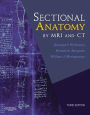
The New Edition of this up-to-date comprehensive sectional anatomy atlas features all new images, demonstrating the latest in MRI technology. It provides carefully labeled MRIs for all body parts, as well as a schematic diagram and concise statements that explain the correlations between the bones and tissues. Three new editors present superior images for abdominal and other difficult areas and offer their expertise in their respective region. View all body images in three standard planes, axial, coronal, and sagittal. Visualize the orientation of every image via schematic diagrams of each body region. Gain a complete understanding of the correlations between body structures.
| Listing 1 - 8 of 8 |
Sort by
|

 Search
 Feedback
 About UniCat
  Help
Help News
News