
 Search
 Feedback
 About UniCat
  Help
Help News
News
| Listing 1 - 10 of 19 | << page >> |
Sort by
|
Book
ISBN: 1596931450 9781596931459 9781596931442 1596931442 Year: 2008 Publisher: Boston ; London : Artech House,
Abstract | Keywords | Export | Availability | Bookmark
 Loading...
Choose an application
- Reference Manager
- EndNote
- RefWorks (Direct export to RefWorks)
This groundbreaking resource offers you exclusive coverage of the latest techniques in diagnostic and therapeutic 3-D ultrasound imaging instrumentation and techniques. Providing a solid overview of potential applications in clinical practice, you find need-to-know details on major diseases, including vascular diseases, breast cancer, cardiac abnormalities and prostate cancer.
Ultrasonic imaging. --- Diagnostic ultrasonic imaging. --- Ultrasonic waves --- Supersonic therapy --- Ultrasonic therapy --- Diagnosis, Ultrasonic --- Diagnostic sonography --- Diagnostic ultrasonics --- Diagnostic ultrasonography --- Diagnostic ultrasound --- Medical diagnostic ultrasonic imaging --- Medical ultrasonography --- Ultrasonic diagnosis --- Ultrasonic diagnostic imaging --- Ultrasonic imaging --- Diagnostic imaging --- Ultrasonics in medicine --- Echography --- Imaging, Ultrasonic --- Sonography --- Ultrasonography --- Acoustic imaging --- Cross-sectional imaging --- Ultrasonics --- Therapeutic use. --- Diagnostic use
Book
ISBN: 1281309958 9786611309954 0387776346 0387776338 Year: 2008 Publisher: New York : Springer,
Abstract | Keywords | Export | Availability | Bookmark
Loading...Choose an application
- Reference Manager
- EndNote
- RefWorks (Direct export to RefWorks)
Thyroid Ultrasound and Ultrasound-Guided FNA, Second Edition is a "user friendly" book for the clinician, using ultrasound in the evaluation and management of thyroid disease. It reviews new information regarding ultrasound and the subtleties one needs to know in the application of this technique. With abundant ultrasound images, it demonstrates how ultrasound is integrated with the patient history, physical exam, and other thyroid tests (especially FNA biopsy) providing information on improving care. The book presents numerous, new innovative uses of ultrasound that are being implemented worldwide. Foreword by Leonard Wartofsky, MD, MACP, Chairman, Department of Medicine, Washington Hospital Center, Washington, DC.
Thyroid gland --- Ultrasonic imaging. --- Needle biopsy. --- Endocrine glands --- Hypothalamic-pituitary-thyroid axis --- Endocrinology. --- Radiology, Medical. --- Diagnosis, Ultrasonic. --- Oncology . --- Otorhinolaryngology. --- Internal medicine. --- Diagnostic Radiology. --- Ultrasound. --- Oncology. --- Internal Medicine. --- Medicine, Internal --- Medicine --- Ear, nose, and throat diseases --- ENT diseases --- Otorhinolaryngology --- Tumors --- Diagnosis, Ultrasonic --- Diagnostic sonography --- Diagnostic ultrasonics --- Diagnostic ultrasonography --- Diagnostic ultrasound --- Medical diagnostic ultrasonic imaging --- Medical ultrasonography --- Ultrasonic diagnosis --- Ultrasonic diagnostic imaging --- Ultrasonic imaging --- Ultrasonic waves --- Diagnostic imaging --- Ultrasonics in medicine --- Clinical radiology --- Radiology, Medical --- Radiology (Medicine) --- Medical physics --- Internal medicine --- Hormones --- Diagnostic use
ISBN: 1281206091 9786611206093 1846285844 1846285208 Year: 2008 Publisher: London : Springer-Verlag London Limited,
Abstract | Keywords | Export | Availability | Bookmark
Loading...Choose an application
- Reference Manager
- EndNote
- RefWorks (Direct export to RefWorks)
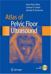
Ultrasound has replaced X-ray as the main imaging modality for the diagnosis of pelvic floor disorders in the female. Most recent developments - volume contrast, speckle reduction and multi-slice imaging - have markedly enhanced spatial resolution and ease of use, with the result that ultrasound now enables cost-effective and non-invasive demonstration of bladder neck and pelvic organ mobility, vaginal, urethral and levator ani function and anatomy, and anorectal anatomy. Atlas of Pelvic Floor Ultrasound provides an introduction to pelvic floor imaging, as well as a resource to be used during initial and more advanced practice. This atlas is an invaluable resource for gynecologists, urogynecologists, female urologists, sonologists, radiologists, and physiotherapists.
Pelvic floor --- Diagnosis --- Ultrasonic imaging --- Pelvic floof --- Floor of pelvis --- Pelvis --- Radiology, Medical. --- Diagnosis, Ultrasonic. --- Urology. --- Gynecology. --- Imaging / Radiology. --- Ultrasound. --- Gynaecology --- Medicine --- Generative organs, Female --- Genitourinary organs --- Diagnosis, Ultrasonic --- Diagnostic sonography --- Diagnostic ultrasonics --- Diagnostic ultrasonography --- Diagnostic ultrasound --- Medical diagnostic ultrasonic imaging --- Medical ultrasonography --- Ultrasonic diagnosis --- Ultrasonic diagnostic imaging --- Ultrasonic waves --- Diagnostic imaging --- Ultrasonics in medicine --- Clinical radiology --- Radiology, Medical --- Radiology (Medicine) --- Medical physics --- Diseases --- Diagnostic use --- Radiology. --- Gynecology . --- Radiological physics --- Physics --- Radiation
Book
ISBN: 128111698X 9786611116989 3540724281 3540724273 3642091490 Year: 2008 Publisher: Berlin : Springer,
Abstract | Keywords | Export | Availability | Bookmark
Loading...Choose an application
- Reference Manager
- EndNote
- RefWorks (Direct export to RefWorks)
Chest sonography is an established procedure in the stepwise imaging diagnosis of pulmonary and pleural disease. It is the method of choice to distinguish between solid and liquid lesions and allows the investigator to make an unequivocal diagnosis without exposing the patient to costly and stressful procedures. This book presents the state of the art in chest investigation by means of ultrasonography. A number of excellent illustrations and the compact text provide concise and easy-to-assimilate information about the diagnostic procedure. Basic elements such as indications, investigation techniques and image artifacts are detailed in separate chapters.
Chest --- Ultrasonic imaging. --- Diseases --- Diagnosis. --- Thorax, Human --- Thorax (Zoology) --- Viscera --- Diagnosis, Ultrasonic. --- Radiology, Medical. --- Pneumology. --- Ultrasound. --- Imaging / Radiology. --- Pneumology/Respiratory System. --- Clinical radiology --- Radiology, Medical --- Radiology (Medicine) --- Medical physics --- Diagnosis, Ultrasonic --- Diagnostic sonography --- Diagnostic ultrasonics --- Diagnostic ultrasonography --- Diagnostic ultrasound --- Medical diagnostic ultrasonic imaging --- Medical ultrasonography --- Ultrasonic diagnosis --- Ultrasonic diagnostic imaging --- Ultrasonic imaging --- Ultrasonic waves --- Diagnostic imaging --- Ultrasonics in medicine --- Diagnostic use --- Radiology. --- Respiratory organs—Diseases. --- Radiological physics --- Physics --- Radiation

ISBN: 9780387681580 0387681582 9780387681597 0387681590 Year: 2008 Publisher: New York, NY : Springer,
Abstract | Keywords | Export | Availability | Bookmark
Loading...Choose an application
- Reference Manager
- EndNote
- RefWorks (Direct export to RefWorks)
There are few situations in anesthesia where precise anatomic location is more important than in regional anesthesia. But, of course, any anesthesiologist who performs regional on a regular basis is fully aware of the frustration of attempting to locate nerves on a trial and error basis. Ultrasound imaging now enables us to visualize nerves, and this exciting technology offers several distinct benefits over conventional nerve locating techniques. The Atlas of Ultrasound and Nerve Stimulation-Guided Regional Anesthesia illustrates how to use ultrasound technology and nerve stimulation techniques to achieve consistently good results. Throughout the book, ultrasound images are correlated with MRI images to enhance anatomic identification. In addition, peripheral nerve block techniques for upper and lower extremities and the trunk are demonstrated step-by-step. With the luxury of being able to actually see the target nerve and the course of the needle as it approaches that nerve, anesthesiologists can now perform regional anesthesia with much greater accuracy. This approach allows the anesthesiologist to conduct regional anesthesia with much greater confidence and efficiency and in doing so the patient is the ultimate beneficiary in terms of success and safety. The book features well-illustrated comparisons of anatomic drawings, cadaveric images, and ultrasound and MRI images. Also: Detailed description of relevant anatomy followed by a clinical description of performing ultrasound imaging and subsequent blockade of target nerves Side-by-side comparison of labeled and unlabeled ultrasound images simulating the clinician’s experience in everyday practice Both common and alternative approaches are discussed in detail, each discussion calling upon the wisdom of experts in the field of regional anesthesia Clinical pearls about needle adjustment included in troubleshooting tables in the nerve stimulation sections.
Anesthesia, Conduction --- Ultrasonography --- Conduction anesthesia --- Nerve block --- Ultrasonics in surgery --- Anesthesiology. --- Radiology. --- Pain medicine. --- Critical care medicine. --- Emergency medicine. --- Ultrasound. --- Pain Medicine. --- Imaging / Radiology. --- Intensive / Critical Care Medicine. --- Emergency Medicine. --- Medicine, Emergency --- Medicine --- Critical care medicine --- Disaster medicine --- Medical emergencies --- Intensive care --- Intensive medicine --- Emergency medicine --- Intensive care units --- Algiatry --- Radiological physics --- Physics --- Radiation --- Anaesthesiology --- Surgery --- Ultrasonics in medicine --- Neural blockade --- Block anesthesia --- Perineural anesthesia --- Regional anesthesia --- Anesthesia
Book
ISBN: 1281871362 9786611871369 1848000901 1848000898 144715858X Year: 2008 Publisher: London : Springer,
Abstract | Keywords | Export | Availability | Bookmark
Loading...Choose an application
- Reference Manager
- EndNote
- RefWorks (Direct export to RefWorks)
In the early 20th century, plain film radiography probably evoked the same sense of wonder that we now associate with cardiac magnetic resonance (CMR). Extensive technical developments and a growth of studies in the literature have increased demand for CMR, but the availability of competing tests and the lack of training opportunities have been limiting. The complexity of CMR examinations and the lack of standardization in protocols between centers likely also hinder its widespread adoption. Cardiovascular MRI in Practice has been written to tackle these issues. This text resource outlines the systematic approach to CMR interpretation. The depiction of a "core exam" and the modifications used for a variety of patient circumstances are demonstrated using simple visual assessment of the images. Special emphasis on the advantages of CMR relative to other modalities reinforces practical learning objectives, organized so that the reader starts with patient images – as one would in a clinical scenario – and works back to the didactic material. This text reference is designed for all cardiologists and cardiovascular radiologists. It is also highly relevant for those in training in order to work through and practice reporting cases using this modality.
Cardiovascular system --- Magnetic resonance imaging. --- Diseases --- Diagnosis. --- Circulatory system --- Vascular system --- Blood --- Circulation --- Cardiology. --- Radiology, Medical. --- Internal medicine. --- Diagnosis, Ultrasonic. --- Heart --- Imaging / Radiology. --- Diagnostic Radiology. --- Internal Medicine. --- Ultrasound. --- Cardiac Surgery. --- Surgery. --- Internal medicine --- Medicine, Internal --- Medicine --- Clinical radiology --- Radiology, Medical --- Radiology (Medicine) --- Medical physics --- Cardiac surgery --- Open-heart surgery --- Diagnosis, Ultrasonic --- Diagnostic sonography --- Diagnostic ultrasonics --- Diagnostic ultrasonography --- Diagnostic ultrasound --- Medical diagnostic ultrasonic imaging --- Medical ultrasonography --- Ultrasonic diagnosis --- Ultrasonic diagnostic imaging --- Ultrasonic imaging --- Ultrasonic waves --- Diagnostic imaging --- Ultrasonics in medicine --- Surgery --- Diagnostic use --- Radiology. --- Cardiac surgery. --- Radiological physics --- Physics --- Radiation

ISBN: 1281397598 9786611397593 3540689176 3540438521 3642078621 Year: 2008 Publisher: Berlin : Springer,
Abstract | Keywords | Export | Availability | Bookmark
Loading...Choose an application
- Reference Manager
- EndNote
- RefWorks (Direct export to RefWorks)
Back cover text Sonography of the gastrointestinal tract in fetuses, neonates and children entails no known biological risk, permits serial scanning and can provide information unobtainable with any other imaging modality. In experienced hands it can be used as the initial imaging technique in a number of gastrointestinal diseases and conditions. This book provides a comprehensive account of the current state of the art regarding sonography in this context. An introductory chapter compares the merits of sonography and magnetic resonance imaging of the fetal gastrointestinal tract. Subsequent chapters focus on the technique, pitfalls and findings in a wide variety of applications, including antropyloric diseases, bowel obstruction, bowel wall thickening, colitis, appendicitis, some types of intussusception, abdominal wall and umbilical abnormalities, intraperitoneal tumors, and trauma. In each case the sonographic morphology is considered in depth with the aid of high-quality illustrations. A concluding chapter comprises a quiz based on 15 case reports. Gastrointestinal Tract Sonography in Fetuses and Children will be of value to all with an interest in this field.
Pediatric gastroenterology --- Gastrointestinal system --- Pediatric diagnostic imaging. --- Fetus --- Diagnosis. --- Ultrasonic imaging. --- Diseases --- Prenatal diagnosis --- Children --- Diagnostic imaging --- Gastro-intestinal system --- Gastrointestinal tract --- GI tract --- Tract, Gastrointestinal --- Tract, GI --- Alimentary canal --- Digestive organs --- Diagnosis --- Radiology, Medical. --- Diagnosis, Ultrasonic. --- Obstetrics. --- Pediatrics. --- Gastroenterology. --- Imaging / Radiology. --- Ultrasound. --- Obstetrics/Perinatology/Midwifery. --- Clinical radiology --- Radiology, Medical --- Radiology (Medicine) --- Medical physics --- Internal medicine --- Paediatrics --- Pediatric medicine --- Medicine --- Maternal-fetal medicine --- Diagnosis, Ultrasonic --- Diagnostic sonography --- Diagnostic ultrasonics --- Diagnostic ultrasonography --- Diagnostic ultrasound --- Medical diagnostic ultrasonic imaging --- Medical ultrasonography --- Ultrasonic diagnosis --- Ultrasonic diagnostic imaging --- Ultrasonic imaging --- Ultrasonic waves --- Ultrasonics in medicine --- Health and hygiene --- Diagnostic use --- Radiology. --- Gastroenterology . --- Radiological physics --- Physics --- Radiation
ISBN: 1281871559 9786611871550 3540490841 3540490833 3642080367 Year: 2008 Publisher: Berlin : Springer,
Abstract | Keywords | Export | Availability | Bookmark
Loading...Choose an application
- Reference Manager
- EndNote
- RefWorks (Direct export to RefWorks)
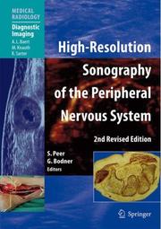
Since the first edition of this book, sonography of the peripheral nervous system has evolved further. Not only has knowledge of the sonographic features of common neurological disorders deepened, but constant research has led to the application of sonography in the diagnosis and guided treatment of disorders that were not previously accessible. This second, revised edition reflects these ongoing developments in various ways: many state of the art high-resolution images are included, the text has been adapted to reflect the current state of the literature, and information is presented using a more modern layout. Anatomic/sonographic correlation is considered in depth, and examination technique with modern sonography equipment is explained. This book provides a practical, clinically oriented overview of all aspects of sonographic diagnosis and interventional therapy of the peripheral nervous system and will be of value to all with an interest in this field.
Nerves, Peripheral --- Ultrasonic imaging. --- Nervous system, Peripheral --- Peripheral nerves --- Peripheral nervous system --- Nervous system --- Radiology, Medical. --- Diagnosis, Ultrasonic. --- Neurology. --- Orthopedic surgery. --- Imaging / Radiology. --- Diagnostic Radiology. --- Ultrasound. --- Neuroradiology. --- Surgical Orthopedics. --- Operative orthopedics --- Orthopedics --- Surgery, Operative --- Medicine --- Neuropsychiatry --- Diagnosis, Ultrasonic --- Diagnostic sonography --- Diagnostic ultrasonics --- Diagnostic ultrasonography --- Diagnostic ultrasound --- Medical diagnostic ultrasonic imaging --- Medical ultrasonography --- Ultrasonic diagnosis --- Ultrasonic diagnostic imaging --- Ultrasonic imaging --- Ultrasonic waves --- Diagnostic imaging --- Ultrasonics in medicine --- Clinical radiology --- Radiology, Medical --- Radiology (Medicine) --- Medical physics --- Diseases --- Diagnostic use --- Radiology. --- Neurology . --- Orthopedics. --- Orthopaedics --- Orthopedia --- Surgery --- Neuroradiography --- Neuroradiology --- Radiological physics --- Physics --- Radiation --- Ultrasonics. --- Radiography. --- Inaudible sound --- Supersonics --- Sound --- Sound pressure
Book
ISBN: 1281871680 9786611871680 3540712070 3540712062 Year: 2008 Publisher: Berlin : Springer,
Abstract | Keywords | Export | Availability | Bookmark
Loading...Choose an application
- Reference Manager
- EndNote
- RefWorks (Direct export to RefWorks)
In my ? rst days as a Radiology resident I remember myself in dire need of a book in which I could begin to study the specialty. In those days I was thirsty for radiolo- cal knowledge and was recommended a classic manual on conventional chest X-ray which was the very same the oldest radiologist of the staff had studied when he was a beginner. Unfortunately the book I was recommended covered less than 5% of the specialty as it is conceived nowadays. “Learning Diagnostic Imaging. A Teaching File” is intended to provide medical students, residents of Radiology, and anybody else beginning to be involved in the radiological world, with a useful tool that gives them a quick and comprehensive overview of Radiology. With this book, written in a user-friendly format, Radiology residents, nurses, technicians, and medical students would see their ? rst radiolo- cal images in a sort of introduction of what will become their professional activities in the rest of their lives.
Diagnosis, Radioscopic --- Diagnosis, Radioscopic. --- Diagnosis, Radiographic --- Radiodiagnosis --- Radioscopic diagnosis --- Roentgenology, Diagnostic --- X-ray diagnosis --- Diagnosis --- Radiography, Medical --- Radiology, Medical. --- Interventional radiology. --- Nuclear medicine. --- Diagnosis, Ultrasonic. --- Diagnostic Radiology. --- Interventional Radiology. --- Imaging / Radiology. --- Nuclear Medicine. --- Ultrasound. --- Diagnosis, Ultrasonic --- Diagnostic sonography --- Diagnostic ultrasonics --- Diagnostic ultrasonography --- Diagnostic ultrasound --- Medical diagnostic ultrasonic imaging --- Medical ultrasonography --- Ultrasonic diagnosis --- Ultrasonic diagnostic imaging --- Ultrasonic imaging --- Ultrasonic waves --- Diagnostic imaging --- Ultrasonics in medicine --- Atomic medicine --- Radioisotopes in medicine --- Medical radiology --- Radioactive tracers --- Radioactivity --- Radiology, Interventional --- Therapeutics --- Clinical radiology --- Radiology, Medical --- Radiology (Medicine) --- Medical physics --- Diagnostic use --- Physiological effect --- Radiology. --- Interventional radiology . --- Radiological physics --- Physics --- Radiation
Book
ISBN: 9788847011113 8847011108 9788847011106 9786612824470 1282824473 8847011116 Year: 2008 Publisher: Milano : Springer-Verlag,
Abstract | Keywords | Export | Availability | Bookmark
Loading...Choose an application
- Reference Manager
- EndNote
- RefWorks (Direct export to RefWorks)
La TC spirale multistrato (TCMS) è una metodica cardine nella diagnostica addominale, in grado di offrire un contributo spesso determinante per l’inquadramento clinico del paziente. Una diagnosi accurata non può, comunque, prescindere da una rigorosa tecnica di esame, in continua evoluzione parallelamente allo sviluppo delle apparecchiature. Attraverso la presentazione di casi riguardanti le principali patologie addominali, il volume rappresenta per il Radiologo generale e per lo Specializzando una guida snella in grado di supportarli nelle decisioni riguardanti l’ottimizzazione dei protocolli di studio in relazione al quesito clinico.
Medicine & Public Health. --- Imaging / Radiology. --- Diagnostic Radiology. --- Ultrasound. --- Medicine. --- Medical radiology --- Diagnosis, Ultrasonic. --- Médecine --- Radiologie médicale --- Abdomen -- Diseases -- Diagnosis. --- Abdomen -- Radiography. --- Abdomen -- Radionuclide imaging. --- Cardiovascular system -- Tomography. --- Radioisotope scanning. --- Spiral computed tomography. --- Medicine --- Health & Biological Sciences --- Radiology, MRI, Ultrasonography & Medical Physics --- Radiology, Medical. --- Clinical radiology --- Radiology, Medical --- Radiology (Medicine) --- Helical computed tomography --- Helical/spiral computed tomography --- Helical/spiral CT (Tomography) --- Spiral CT (Tomography) --- Radiology. --- Medical physics --- Tomography --- Diagnosis, Ultrasonic --- Diagnostic sonography --- Diagnostic ultrasonics --- Diagnostic ultrasonography --- Diagnostic ultrasound --- Medical diagnostic ultrasonic imaging --- Medical ultrasonography --- Ultrasonic diagnosis --- Ultrasonic diagnostic imaging --- Ultrasonic imaging --- Ultrasonic waves --- Diagnostic imaging --- Ultrasonics in medicine --- Diagnostic use --- Radiological physics --- Physics --- Radiation --- Medical radiology.
| Listing 1 - 10 of 19 | << page >> |
Sort by
|