| Listing 1 - 10 of 10 |
Sort by
|
Book
Abstract | Keywords | Export | Availability | Bookmark
 Loading...
Choose an application
- Reference Manager
- EndNote
- RefWorks (Direct export to RefWorks)
Magnetic Resonance Imaging --- Tomography, X-Ray Computed --- Anatomy, Cross-Sectional --- Thorax --- Abdomen --- Pelvis
Book
Year: 2007 Publisher: [Place of publication not identified] Churchill Livingstone Elsevier
Abstract | Keywords | Export | Availability | Bookmark
 Loading...
Choose an application
- Reference Manager
- EndNote
- RefWorks (Direct export to RefWorks)
Human anatomy --- Magnetic resonance imaging --- Tomography --- Anatomy, Regional. --- Magnetic Resonance Imaging. --- Tomography, X-Ray Computed.
Dissertation
ISBN: 9789036730617 Year: 2007 Publisher: Groningen Grafimedia.
Abstract | Keywords | Export | Availability | Bookmark
 Loading...
Choose an application
- Reference Manager
- EndNote
- RefWorks (Direct export to RefWorks)
Lung Neoplasms --- Lung Neoplasms --- Tomography, X-Ray Computed --- diagnosis --- radiography --- methods
ISBN: 9781588905666 1588905667 Year: 2007 Publisher: Stuttgart: Thieme,
Abstract | Keywords | Export | Availability | Bookmark
 Loading...
Choose an application
- Reference Manager
- EndNote
- RefWorks (Direct export to RefWorks)
Magnetic Resonance Imaging --- Tomography, X-Ray Computed --- Anatomy, Cross-Sectional --- Spine --- Extremities --- Joints --- Human anatomy --- Tomography --- Magnetic resonance imaging
ISBN: 0781757657 9780781757652 Year: 2007 Publisher: Philadelphia : Wolters Kluwer/Lippincott Williams & Wilkins,
Abstract | Keywords | Export | Availability | Bookmark
 Loading...
Choose an application
- Reference Manager
- EndNote
- RefWorks (Direct export to RefWorks)
The thoroughly revised, updated Fourth Edition of this classic reference provides authoritative, current guidelines on chest imaging using state-of-the-art technologies, including multidetector CT, MRI, PET, and integrated CT-PET scanning. This edition features a brand-new chapter on cardiac imaging. Extensive descriptions of the use of PET have been added to the chapters on lung cancer, focal lung disease, and the pleura, chest wall, and diaphragm. Also included are recent PIOPED II findings on the role of CT angiography and CT venography in detecting pulmonary embolism. Complementing the text are 2,300 CT, MR, and PET scans made on the latest-generation scanners.
Chest --- Chest --- Magnetic Resonance Imaging. --- Radiography, Thoracic. --- Thorax --- Tomography, X-Ray Computed. --- Magnetic resonance imaging. --- Tomography. --- pathology.
ISBN: 9781841846255 1841846252 Year: 2007 Publisher: London : Informa Healthcare,
Abstract | Keywords | Export | Availability | Bookmark
 Loading...
Choose an application
- Reference Manager
- EndNote
- RefWorks (Direct export to RefWorks)
Cardiovascular system --- Cardiovascular Diseases --- Coronary Angiography --- Tomography, X-Ray Computed --- Appareil cardiovasculaire --- Tomography --- radiography. --- methods. --- methods. --- Tomographie

ISBN: 158890475X 313125503X 9781588904751 9783131255037 Year: 2007 Publisher: Stuttgart : Thieme,
Abstract | Keywords | Export | Availability | Bookmark
 Loading...
Choose an application
- Reference Manager
- EndNote
- RefWorks (Direct export to RefWorks)
Anatomy, Regional --- Human anatomy --- Magnetic Resonance Imaging --- Magnetic resonance imaging --- Tomography --- Tomography, X-Ray Computed --- atlases. --- Anatomy, Cross-Sectional --- Head --- Neck --- Spine --- Joints
ISBN: 9781931884426 1931884420 9781931884433 1931884439 Year: 2007 Publisher: Canada Amirsys
Abstract | Keywords | Export | Availability | Bookmark
 Loading...
Choose an application
- Reference Manager
- EndNote
- RefWorks (Direct export to RefWorks)
FThis volume of the landmark Diagnostic and Surgical Imaging Anatomy series combines a rich pictorial database of high-resolution images and lavish, 3-D color illustrations to help you interpret multiplanar scans with confidence. The book brings you close up to see key structures with meticulously labeled anatomic landmarks from axial, coronal, and sagittal planes. Contents include over 150 detail-revealing 3-D color illustrations, over 950 high-resolution digital scans, and at-a-glance imaging summaries for the knee, ankle, and foot.
Ankle --- Foot --- Knee --- Magnetic Resonance Spectroscopy --- Tomography, X-Ray Computed --- Anatomy --- Imaging --- anatomy & histology --- Genou --- Cheville --- Pied --- Atlases. --- Anatomie --- Atlas --- Imagerie --- Diagnose --- Chirurgie --- Knieën --- Enkeltrauma --- Voeten --- Anatomy & histology --- Knie --- VOET --- Breuk (geneeskunde) --- Letsel --- Orthese --- Wervelkolom --- Voet --- Diagnostiek
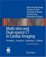
ISBN: 1280727098 9786610727094 3540495460 3540255230 Year: 2007 Publisher: Berlin Springer
Abstract | Keywords | Export | Availability | Bookmark
 Loading...
Choose an application
- Reference Manager
- EndNote
- RefWorks (Direct export to RefWorks)
Cardiac diseases, and in particular coronary artery disease, are the leading cause of death and morbidity in industrialized countries. The development of non-invasive imaging techniques for the heart and the coronary arteries has been considered a key element in improving patient care. A breakthrough in cardiac imaging using CT occurred in 1998, with the introduction of multi-slice computed tomography (CT). Since then, amazing advances in performance have taken place with scanners that acquire up to 64 slices per rotation. This book discusses the state-of-the-art developments in multi-slice CT for cardiac imaging as well as those that can be anticipated in the future. It serves as a comprehensive work that covers all aspects of this technology, from the technical fundamentals and image evaluation all the way to clinical indications and protocol recommendations. This fully reworked second edition draws on the most recent clinical experience obtained with 16- and 64-slice CT scanners by world-leading experts from Europe and the United States. It also includes "hands-on" experience in the form of 10 representative clinical case studies, which are included on the accompanying CD. As a further highlight, the latest results of the very recently introduced dual-source CT, which may soon represent the CT technology of choice for cardiac applications, are presented. This book will not only convince the reader that multi-slice cardiac CT has arrived in clinical practice, it will also make a significant contribution to the education of radiologists, cardiologists, technologists, and physicistswhether newcomers, experienced users, or researchers.
Heart --- Imaging. --- Diseases --- Diagnosis. --- Cardiac diagnostic imaging --- Cardiac imaging --- Diagnostic cardiac imaging --- Imaging of the heart --- Imaging --- WG 141.5 Cardiovascular Diseases, Diagnosis and Therapeutics -- Specific diagnostic methods --- Diagnosis --- Heart Diseases --- Tomography, X-Ray Computed --- Radiology, Medical. --- Cardiology. --- Imaging / Radiology. --- Internal medicine --- Clinical radiology --- Radiology, Medical --- Radiology (Medicine) --- Medical physics --- Radiology. --- Radiological physics --- Physics --- Radiation

ISBN: 1281148288 9786611148287 0387392580 0387392548 9780387392547 9780387392585 Year: 2007 Publisher: Boston, MA Springer Science+Business Media, LLC
Abstract | Keywords | Export | Availability | Bookmark
 Loading...
Choose an application
- Reference Manager
- EndNote
- RefWorks (Direct export to RefWorks)
Micro-Tomographic Atlas of the Mouse Skeleton Professor Itai Bab, Chief, Bone Laboratory, The Hebrew University of Jerusalem, Jerusalem, Israel Professor Ralph Müller, Director, Center for Bioengineering Research and Education, ETH Zürich, Switzerland Micro-Tomographic Atlas of the Mouse Skeleton serves as an essential guide containing unique systematic description of all calcified components of the mouse. This detailed atlas fulfils an emerging need for high resolution anatomical details as mice become a standard laboratory animal in skeletal research and the use of m CT technology is rapidly increasing as a key analytical tool in the study of bone. Key Features: Includes over 200 high resolution, two- and three dimensional m CT images of the exterior and interiors of all bones and joints Offers the spatial relationship of individual bones within complex skeletal units (e.g., skull, thorax, pelvis, extremities). All images are accompanied by detailed explanatory text that highlights special features and newly reported structures. Available for the first time in the Atlas: Detailed information on the micro-anatomy of the murine skeleton essential for the design of experiments and interpretation of results Comparative analyses on m CT-based morphometric parameters at the whole bone, cortical and trabecular levels including: Age differences (4-40 weeks) Gender differences Differences between main mouse strains (C57Bl/6J, SJL, C3H) Micro-Tomographic Atlas of the Mouse Skeleton offers a practical, comprehensive desk reference for all scientists and students interested in skeletal biology.
Mice --- Skeleton --- Anatomy --- Tomography. --- House mice --- House mouse --- Mouse --- Mus musculus --- Rodents --- Osteology --- Bones --- Bone and Bones --- Tomography, X-Ray Computed --- radiography --- anatomy & histology --- Biomedical engineering. --- Zoology. --- Animal physiology. --- Human anatomy. --- Neurobiology. --- Developmental biology. --- Biomedical Engineering and Bioengineering. --- Animal Physiology. --- Anatomy. --- Developmental Biology. --- Development (Biology) --- Biology --- Growth --- Ontogeny --- Neurosciences --- Anatomy, Human --- Human biology --- Medical sciences --- Human body --- Animal physiology --- Animals --- Natural history --- Clinical engineering --- Medical engineering --- Bioengineering --- Biophysics --- Engineering --- Medicine --- Physiology --- Radiography --- Anatomy & histology
| Listing 1 - 10 of 10 |
Sort by
|

 Search
 Feedback
 About UniCat
  Help
Help News
News