| Listing 1 - 10 of 14 | << page >> |
Sort by
|
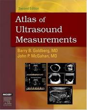
ISBN: 032303229X 9780323032292 Year: 2006 Publisher: Philadelphia (Pa.) : Mosby,
Abstract | Keywords | Export | Availability | Bookmark
 Loading...
Choose an application
- Reference Manager
- EndNote
- RefWorks (Direct export to RefWorks)
Diagnosis, Ultrasonic --- Diagnostic ultrasonic imaging --- Ultrasonic imaging --- Ultrasonography
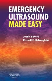
ISBN: 0443101507 Year: 2006 Publisher: Edinburgh ; New York : Elsevier/Churchill Livingstone,
Abstract | Keywords | Export | Availability | Bookmark
 Loading...
Choose an application
- Reference Manager
- EndNote
- RefWorks (Direct export to RefWorks)
Diagnosis, Ultrasonic. --- Diagnostic ultrasonic imaging. --- Emergencies. --- Emergency Medical Services --- Emergency medicine --- Ultrasonic imaging. --- Ultrasonography --- Methods. --- Diagnosis. --- Methods.
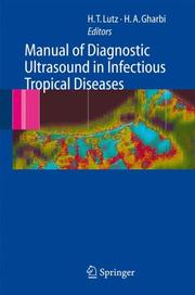
ISBN: 1280412046 9786610412044 3540299505 3540244468 Year: 2006 Publisher: Berlin ; New York : Springer,
Abstract | Keywords | Export | Availability | Bookmark
 Loading...
Choose an application
- Reference Manager
- EndNote
- RefWorks (Direct export to RefWorks)
This manual is intended to fill a gap in the range of books on ultrasound diagnosis, concentrating exclusively on the diagnosis of infectious and tropical diseases. The book starts with a short introduction to ultrasound diagnosis. The main part describes and discusses the organ-related changes that can be detected with ultrasound in infectious and inflammatory diseases. The infectious and parasitic diseases that can successfully be diagnosed with ultrasound are presented in more detail. The important potential of interventional ultrasound diagnosis and therapy in infectious diseases is described. This richly illustrated volume has been written by leading experts from all over the world. It is primarily aimed at users of ultrasound diagnostic instruments in tropical and subtropical countries but will be valuable for physicians anywhere in the world confronted with these diseases.
Diagnostic ultrasonic imaging. --- Tropical medicine. --- Diseases, Tropical --- Hygiene, Tropical --- Medicine --- Public health, Tropical --- Sanitation, Tropical --- Tropical diseases --- Medical climatology --- Diagnosis, Ultrasonic --- Diagnostic sonography --- Diagnostic ultrasonics --- Diagnostic ultrasonography --- Diagnostic ultrasound --- Medical diagnostic ultrasonic imaging --- Medical ultrasonography --- Ultrasonic diagnosis --- Ultrasonic diagnostic imaging --- Ultrasonic imaging --- Ultrasonic waves --- Diagnostic imaging --- Ultrasonics in medicine --- Diagnostic use --- Internal medicine. --- Diagnosis, Ultrasonic. --- Internal Medicine. --- Ultrasound. --- Tropical Medicine. --- Medicine, Internal --- Radiology. --- Radiological physics --- Physics --- Radiation
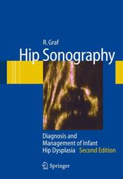
ISBN: 1280635037 9786610635030 3540309586 3540309578 Year: 2006 Publisher: Berlin ; New York : Springer,
Abstract | Keywords | Export | Availability | Bookmark
 Loading...
Choose an application
- Reference Manager
- EndNote
- RefWorks (Direct export to RefWorks)
Sonography of baby hips for the diagnosis of DDH and dysplasia has grown steadily in importance in recent years. A strict standardized technique for investigation of the baby and interpretation of the sonograms has made hip ultrasound reproducible, reliable and independent of examiner skill and experience. Graf’s technique is now used worldwide, and selective or even general screening programmes for all babies are established in many European countries today. The first part of this book includes the fundamentals of hip sonography, static as well as dynamic techniques, anatomical identification of the echograms, typing, a measurement technique and usability check. The second part is an atlas including a summary of the essential data and demonstrating correct and incorrect sonograms in different variations. The book is indispensable for everyone dealing with DDH problems in diagnosis and therapy.
Hip joint --- Infants --- Dislocation, Congenital --- Ultrasonic imaging. --- Coxa --- Joints --- Babies --- Infancy --- Children --- Diagnosis, Ultrasonic. --- Orthopedics. --- Pediatrics. --- Ultrasound. --- Paediatrics --- Pediatric medicine --- Medicine --- Orthopaedics --- Orthopedia --- Surgery --- Diagnosis, Ultrasonic --- Diagnostic sonography --- Diagnostic ultrasonics --- Diagnostic ultrasonography --- Diagnostic ultrasound --- Medical diagnostic ultrasonic imaging --- Medical ultrasonography --- Ultrasonic diagnosis --- Ultrasonic diagnostic imaging --- Ultrasonic imaging --- Ultrasonic waves --- Diagnostic imaging --- Ultrasonics in medicine --- Diseases --- Health and hygiene --- Diagnostic use --- Radiology. --- Radiological physics --- Physics --- Radiation

ISBN: 1280626100 9786610626106 8847005078 8847003369 Year: 2006 Publisher: Milan ; New York : Springer,
Abstract | Keywords | Export | Availability | Bookmark
 Loading...
Choose an application
- Reference Manager
- EndNote
- RefWorks (Direct export to RefWorks)
New three-dimensional endoanal and endorectal ultrasonographic and magnetic resonance imaging techniques have given better insight into the complex anatomy of the pelvic floor and its pathologic modification in benign anorectal diseases. Obstetrical events leading to fecal incontinence in females, the relationship between fistulous tracks and the sphincter complex, and mechanisms of obstructed defecation syndrome can now be accurately evaluated, which is of fundamental importance for decision making. Thanks to improvements in the diagnosis of these disorders, new forms of treatment have been developed with better outcome for patients. This book is aimed at general and colorectal surgeons, radiologists, gastroenterologists and gynecologists with a special interest in this field. It is also relevant to everyone who wants to improve their understanding of the fundamental principles of pelvic floor disorders.
Rectum --- Gastrointestinal system --- Diseases. --- Proctology --- Abdomen --- Gastroenterology. --- Diagnosis, Ultrasonic. --- Colon (Anatomy) --- Surgery. --- Gynecology. --- Abdominal Surgery. --- Ultrasound. --- Colorectal Surgery. --- Gynaecology --- Medicine --- Generative organs, Female --- Surgery, Primitive --- Diagnosis, Ultrasonic --- Diagnostic sonography --- Diagnostic ultrasonics --- Diagnostic ultrasonography --- Diagnostic ultrasound --- Medical diagnostic ultrasonic imaging --- Medical ultrasonography --- Ultrasonic diagnosis --- Ultrasonic diagnostic imaging --- Ultrasonic imaging --- Ultrasonic waves --- Diagnostic imaging --- Ultrasonics in medicine --- Internal medicine --- Digestive organs --- Abdominal surgery --- Laparotomy --- Diseases --- Diagnostic use --- Abdominal surgery. --- Gastroenterology . --- Radiology. --- Rectum—Surgery . --- Gynecology . --- Radiological physics --- Physics --- Radiation

ISBN: 1280626135 9786610626137 8847004586 8847004578 Year: 2006 Publisher: Milan ; Berlin : Springer,
Abstract | Keywords | Export | Availability | Bookmark
 Loading...
Choose an application
- Reference Manager
- EndNote
- RefWorks (Direct export to RefWorks)
In recent years, the imaging-based diagnosis of mass liver lesions has become increasingly complex due to the number and morphological variability of lesions that modern imaging techniques are currently able to display. If the sensitivity in detection has greatly increased, characterisation has remained difficult and represents a critical challenge for the clinician. The availability of blood-pool contrast agents for ultrasound (US), in particular second-generation US contrast agents based on perfluorocarbon- or sulfur-hexafluoride-filled microbubbles, and the development of contrast-specific software and technologies have opened up new perspectives both for the immediate characterisation of any mass lesion detected in the liver and for increasing the sensitivity of US in the detection of liver metastases. Taking into account the great impact of this new technology on clinical practice, the European Federation of Societies for Ultrasound in Medicine and Biology (EFSUMB) organised, in January 2004, in Rotterdam, a consensus meeting of experts in order to develop guidelines for the use of US contrast agents in the diagnosis of liver diseases . These guidelines, as well as discussions of further advances in the clinical application of contrast-enhanced harmonic US are presented in this book by an internationally renowned group of experts. The book represents provides an important starting point for clinical implementation of this new diagnostic procedure.
Diagnostic imaging. --- Liver --- Treatment. --- Abdomen --- Biliary tract --- Clinical imaging --- Imaging, Diagnostic --- Medical diagnostic imaging --- Medical imaging --- Noninvasive medical imaging --- Diagnosis, Noninvasive --- Imaging systems in medicine --- Radiology, Medical. --- Diagnosis, Ultrasonic. --- Imaging / Radiology. --- Ultrasound. --- Diagnostic Radiology. --- Diagnosis, Ultrasonic --- Diagnostic sonography --- Diagnostic ultrasonics --- Diagnostic ultrasonography --- Diagnostic ultrasound --- Medical diagnostic ultrasonic imaging --- Medical ultrasonography --- Ultrasonic diagnosis --- Ultrasonic diagnostic imaging --- Ultrasonic imaging --- Ultrasonic waves --- Diagnostic imaging --- Ultrasonics in medicine --- Clinical radiology --- Radiology, Medical --- Radiology (Medicine) --- Medical physics --- Diagnostic use --- Radiology. --- Radiological physics --- Physics --- Radiation
Book
ISBN: 1846281466 Year: 2006 Publisher: London : Springer,
Abstract | Keywords | Export | Availability | Bookmark
 Loading...
Choose an application
- Reference Manager
- EndNote
- RefWorks (Direct export to RefWorks)
CT has long been considered an accurate technique in the assessment of cardiac structure and function, but advances in computing power and scanning technology have resulted in its increasing popularity. It is particularly useful in evaluating the myocardium, coronary arteries, pulmonary veins, thoracic aorta, pericardium, and cardiac masses, such as thrombus of the left atrial appendage. Given this wide array of possible diagnoses and the speed at which scans can be performed, it is becoming even more attractive as an advanced, cost-effective and integral part of patient evaluation. Given this growing importance to the practising cardiologist worldwide, this book collates all relevant imaging findings of the use of cardiac CT and presents them in a clinically relevant and practical manner appropriate for residents and fellows in both cardiology and radiology. The images have been supplied by an internationally renowned set of contributing authors and represent the full spectrum of cardiac CT imaging. As ever increasing numbers of clinicians have access to cardiac CT scanners, this book provides all the relevant information to those wanting to diagnose patients using this modality.
Heart --- Cardiovascular system --- Tomography. --- Diseases --- Diagnosis. --- Cardiographic tomography --- Tomography --- Radiography --- Cardiology. --- Radiology, Medical. --- Internal medicine. --- Diagnosis, Ultrasonic. --- Imaging / Radiology. --- Diagnostic Radiology. --- Internal Medicine. --- Ultrasound. --- Cardiac Surgery. --- Surgery. --- Cardiac surgery --- Open-heart surgery --- Diagnosis, Ultrasonic --- Diagnostic sonography --- Diagnostic ultrasonics --- Diagnostic ultrasonography --- Diagnostic ultrasound --- Medical diagnostic ultrasonic imaging --- Medical ultrasonography --- Ultrasonic diagnosis --- Ultrasonic diagnostic imaging --- Ultrasonic imaging --- Ultrasonic waves --- Diagnostic imaging --- Ultrasonics in medicine --- Medicine, Internal --- Medicine --- Clinical radiology --- Radiology, Medical --- Radiology (Medicine) --- Medical physics --- Internal medicine --- Surgery --- Diagnostic use --- Radiology. --- Cardiac surgery. --- Radiological physics --- Physics --- Radiation
ISBN: 1280626119 9786610626113 8847003679 8847003660 Year: 2006 Publisher: Milan ; Berlin : Springer,
Abstract | Keywords | Export | Availability | Bookmark
 Loading...
Choose an application
- Reference Manager
- EndNote
- RefWorks (Direct export to RefWorks)
2-D and 3-D anal ultrasound are among the most recent and advanced tools available for both the diagnosis and the management of anorectal diseases. They are neither expensive nor harmful for the patients and progressively replaced anal mapping with EMG electrodes for the diagnosis of sphincter's defects and anismus, which represents nearly 50% of the cases of chronic constipation. Anal US may provide the clinician with useful information for both classification, diagnosis and management of anorectal sepsis, anal incontinence and anorectal-perineal chronic pain. Almost any case presented in this Atlas shows both imaging and clinical pictures, thus allowing both the radiologist and the clinician to assess the reliability of the exam and the outcome of the selected treatment.
Rectum --- Proctology. --- Diseases. --- Gastroenterology --- Proctology --- Colon (Anatomy) --- Pathology. --- Radiology, Medical. --- Gynecology. --- Diagnosis, Ultrasonic. --- Gastroenterology. --- Colorectal Surgery. --- Imaging / Radiology. --- Ultrasound. --- Surgery. --- Gynaecology --- Medicine --- Generative organs, Female --- Clinical radiology --- Radiology, Medical --- Radiology (Medicine) --- Medical physics --- Disease (Pathology) --- Medical sciences --- Diseases --- Medicine, Preventive --- Internal medicine --- Digestive organs --- Diagnosis, Ultrasonic --- Diagnostic sonography --- Diagnostic ultrasonics --- Diagnostic ultrasonography --- Diagnostic ultrasound --- Medical diagnostic ultrasonic imaging --- Medical ultrasonography --- Ultrasonic diagnosis --- Ultrasonic diagnostic imaging --- Ultrasonic imaging --- Ultrasonic waves --- Diagnostic imaging --- Ultrasonics in medicine --- Diagnostic use --- Rectum—Surgery . --- Radiology. --- Gynecology . --- Gastroenterology . --- Radiological physics --- Physics --- Radiation
ISBN: 1281350354 9786611350352 8847004608 8847004594 Year: 2006 Publisher: Milano : Springer,
Abstract | Keywords | Export | Availability | Bookmark
 Loading...
Choose an application
- Reference Manager
- EndNote
- RefWorks (Direct export to RefWorks)
Questo atlante è ispirato ad una strategia didattica esemplare e intende fornire agli specialisti, coinvolti nella gestione della malattia cardiaca, strumenti comuni per la comprensione ed il trattamento delle varie problematiche ad essa legate nella pratica clinica quotidiana. L’informazione viene selezionata accuratamente e l’ecocardiografia è immersa nel contesto clinico, anatomico, fisiopatologico , terapeutico e chirurgico entro cui è abitualmente utilizzata. È un libro figlio dell’esperienza e della cultura del Brompton Hospital di Londra – dove con Derek Gibson è nata negli anni ’70 l’ecocardiografia moderna, in un ambiente di eccellenza, dove ogni giorno l’attendibilità dell’informazione ecocardiografica veniva tarata sulla spietata verifica clinica, emodinamica, cardiochirurgica. Di Gibson, Michael Henein è oggi il degno successore a capo del più rinomato laboratorio di ecocardiografia del Regno Unito. È un laboratorio gestito da cardiologi clinici che si servono dell’ecocardiografia non come un fine ma come un mezzo (prezioso, prodigiosamente versatile, insostituibile – ma sempre e solo un mezzo) per curare meglio i pazienti.
Echocardiography. --- Cardiology. --- Diagnostic ultrasonic imaging. --- Heart --- Internal medicine --- Diseases --- Diagnosis, Ultrasonic --- Diagnostic sonography --- Diagnostic ultrasonics --- Diagnostic ultrasonography --- Diagnostic ultrasound --- Medical diagnostic ultrasonic imaging --- Medical ultrasonography --- Ultrasonic diagnosis --- Ultrasonic diagnostic imaging --- Ultrasonic imaging --- Ultrasonic waves --- Diagnostic imaging --- Ultrasonics in medicine --- Echo cardiography --- Ultrasonic cardiography --- Ultrasound cardiography --- Cardiography --- Diagnostic ultrasonic imaging --- Diagnostic use --- Imaging --- Radiology, Medical. --- Diagnosis, Ultrasonic. --- Angiography. --- Imaging / Radiology. --- Diagnostic Radiology. --- Ultrasound. --- Angiology. --- Cardiac Surgery. --- Surgery. --- Cardiac surgery --- Open-heart surgery --- Blood-vessels --- Diagnosis, Radioscopic --- Radiography, Medical --- Clinical radiology --- Radiology, Medical --- Radiology (Medicine) --- Medical physics --- Surgery --- Radiography --- Radiology. --- Cardiac surgery. --- Radiological physics --- Physics --- Radiation

ISBN: 1280615826 9786610615827 8847004764 8847004756 Year: 2006 Publisher: New York : Springer,
Abstract | Keywords | Export | Availability | Bookmark
 Loading...
Choose an application
- Reference Manager
- EndNote
- RefWorks (Direct export to RefWorks)
The introduction of microbubble contrast agents and the development of contrast-specific scanning techniques have opened new prospects in ultrasound. The advent of second-generation agents – that enable real-time contrast-enhanced imaging – has been instrumental in improving the acceptance and the reproducibility of examinations. Contrast ultrasound substantially improves detection and characterization of focal liver lesions with respect to baseline studies, and has already been introduced in international guidelines for the diagnosis of liver tumors. The role of contrast agents in vascular ultrasound is also established, and several new clinical applications are emerging. This book, written by the leading experts in the field, provides an up-to-date overview on the clinical value of contrast agents in ultrasound. The volume moves from a background section on technique and methodology to the main sections on the clinical application of contrast ultrasound in the liver and in vascular diseases. A final section discusses results and prospects of contrast ultrasound modality in the other fields.
Ultrasonic imaging. --- Diagnostic ultrasonic imaging. --- Diagnosis, Ultrasonic --- Diagnostic sonography --- Diagnostic ultrasonics --- Diagnostic ultrasonography --- Diagnostic ultrasound --- Medical diagnostic ultrasonic imaging --- Medical ultrasonography --- Ultrasonic diagnosis --- Ultrasonic diagnostic imaging --- Ultrasonic imaging --- Ultrasonic waves --- Diagnostic imaging --- Ultrasonics in medicine --- Echography --- Imaging, Ultrasonic --- Sonography --- Ultrasonography --- Acoustic imaging --- Cross-sectional imaging --- Ultrasonics --- Diagnostic use --- Diagnosis, Ultrasonic. --- Radiology, Medical. --- Clinical medicine. --- Oncology . --- Urology. --- Ultrasound. --- Imaging / Radiology. --- Hepatology. --- Oncology. --- Diagnostic Radiology. --- Medicine --- Genitourinary organs --- Tumors --- Medicine, Clinical --- Clinical radiology --- Radiology, Medical --- Radiology (Medicine) --- Medical physics --- Diseases --- Radiology. --- Gastroenterology --- Radiological physics --- Physics --- Radiation
| Listing 1 - 10 of 14 | << page >> |
Sort by
|

 Search
 Feedback
 About UniCat
  Help
Help News
News