| Listing 1 - 10 of 78 | << page >> |
Sort by
|
Book
ISBN: 9535167049 9533078243 Year: 2012 Publisher: IntechOpen
Abstract | Keywords | Export | Availability | Bookmark
 Loading...
Choose an application
- Reference Manager
- EndNote
- RefWorks (Direct export to RefWorks)
This book's stated purpose is to provide a discussion of the technical basis and clinical applications of positron emission tomography (PET), as well as their recent progress in nuclear medicine. It also summarizes current literature about research and clinical science in PET. The book is divided into two broad sections: basic science and clinical science. The basic science section examines PET imaging processing, kinetic modeling, free software, and radiopharmaceuticals. The clinical science section demonstrates various clinical applications and diagnoses. The text is intended not only for scientists, but also for all clinicians seeking recent information regarding PET.
Tomography, Emission. --- Computerized emission tomography --- Emission tomography --- PET (Tomography) --- PET-CT (Tomography) --- Positron emission tomography --- Positron emission transaxial tomography --- Radionuclide tomography --- Scintigraphy, Tomographic --- Tomography, Radionuclide --- Diagnosis --- Diagnostic imaging --- Positrons --- Radioisotope scanning --- Data processing --- Emission --- Radiology

ISBN: 1281140007 9786611140007 038738295X 0387352759 Year: 2007 Publisher: New York, NY : Springer Science+Business Media, LLC,
Abstract | Keywords | Export | Availability | Bookmark
 Loading...
Choose an application
- Reference Manager
- EndNote
- RefWorks (Direct export to RefWorks)
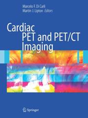
Cardiac PET and hybrid PET/CT have revolutionized the field of cardiovascular imaging and offer new options for diagnosis and management of patients with cardiac conditions. Cardiologists, radiologists, and nuclear medicine specialists must have an up-to-date reference in this rapidly changing field in order to most effectively utilize new technology in practice. Cardiac PET and PET/CT Imaging takes a step-by-step approach to PET and PET/CT instrumentation, imaging, and protocols. Beginning with imaging fundamentals and progressing through advanced discussion of specific applications, this book is a comprehensive source of cardiac PET and PET/CT information. Sections written by and for technologists as well as discussion of MRI and other imaging modalities add to the well-rounded scope of this book. A library of original cases and several hundred illustrations, many in color, complete the text by highlighting interpretation and technical challenges in cardiac PET and PET/CT.
Heart --- Tomography, Emission. --- Imaging. --- Computerized emission tomography --- Emission tomography --- PET (Tomography) --- PET-CT (Tomography) --- Positron emission tomography --- Positron emission transaxial tomography --- Radionuclide tomography --- Scintigraphy, Tomographic --- Tomography, Radionuclide --- Diagnosis --- Diagnostic imaging --- Positrons --- Radioisotope scanning --- Cardiac diagnostic imaging --- Cardiac imaging --- Diagnostic cardiac imaging --- Imaging of the heart --- Data processing --- Emission --- Diseases --- Imaging

ISBN: 079235091X 9401060975 9401149968 Year: 1998 Volume: 51 Publisher: Dordrecht : Kluwer,
Abstract | Keywords | Export | Availability | Bookmark
 Loading...
Choose an application
- Reference Manager
- EndNote
- RefWorks (Direct export to RefWorks)
A critical summary of the state of the art of PET technology and related disciplines (camera physics, radiochemistry, radiopharmacology, computerised brain atlases and databases, etc.), plus a survey of the most recent developments in the clinical and neuroscience applications of PET. The chapters systematically guide the reader from the basics of the technique - including new camera designs, new drugs, and new statistical and image processing approaches - through its most important clinical applications - in neurology, psychiatry and oncology - to the most sophisticated neuroscience applications, including research into human sensory and motor systems, the functional organisation of imagery, volition, attention and consciousness in the human brain.
Radiology. --- Neurosciences. --- Neurology . --- Oncology . --- Imaging / Radiology. --- Neurology. --- Oncology. --- Tumors --- Medicine --- Nervous system --- Neuropsychiatry --- Neural sciences --- Neurological sciences --- Neuroscience --- Medical sciences --- Radiological physics --- Physics --- Radiation --- Diseases --- Tomography, Emission. --- Computerized emission tomography --- Emission tomography --- PET (Tomography) --- PET-CT (Tomography) --- Positron emission tomography --- Positron emission transaxial tomography --- Radionuclide tomography --- Scintigraphy, Tomographic --- Tomography, Radionuclide --- Diagnosis --- Diagnostic imaging --- Positrons --- Radioisotope scanning --- Data processing --- Emission --- Diagnosis. --- Positron-emission tomography
Book
ISBN: 3319548379 3319548360 Year: 2017 Publisher: Cham : Springer International Publishing : Imprint: Springer,
Abstract | Keywords | Export | Availability | Bookmark
 Loading...
Choose an application
- Reference Manager
- EndNote
- RefWorks (Direct export to RefWorks)
This book is a pocket guide to the science and practice of PET/CT imaging of colorectal malignancies. The role of PET/CT in these patients, the characteristic PET/CT findings, and the advantages and limitations of this hybrid modality are all clearly described. In addition, information is provided on clinical presentation, diagnosis, staging, pathology, management, and radiological imaging. The book is published within the Springer series Clinicians' Guides to Radionuclide Hybrid Imaging, which is aimed at nuclear medicine/radiology residents and physicians, referring clinicians, radiographers/technologists, and nurses who routinely work in nuclear medicine and participate in multidisciplinary meetings. Compiled under the auspices of the British Nuclear Medicine Society, the series is the joint work of many colleagues and professionals worldwide who share a common vision and purpose in promoting and supporting nuclear medicine as an important imaging specialty for the diagnosis and management of oncological and non-oncological conditions.
Medicine. --- Nuclear medicine. --- Oncology. --- Medicine & Public Health. --- Nuclear Medicine. --- Tomography, Emission. --- Cancer --- Colon (Anatomy) --- Diagnosis. --- Treatment. --- Computerized emission tomography --- Emission tomography --- PET (Tomography) --- PET-CT (Tomography) --- Positron emission tomography --- Positron emission transaxial tomography --- Radionuclide tomography --- Scintigraphy, Tomographic --- Tomography, Radionuclide --- Diagnosis --- Diagnostic imaging --- Positrons --- Radioisotope scanning --- Data processing --- Emission --- Oncology . --- Tumors --- Atomic medicine --- Radioisotopes in medicine --- Medical radiology --- Radioactive tracers --- Radioactivity --- Physiological effect
Book
ISBN: 3319605070 3319605062 Year: 2018 Publisher: Cham : Springer International Publishing : Imprint: Springer,
Abstract | Keywords | Export | Availability | Bookmark
 Loading...
Choose an application
- Reference Manager
- EndNote
- RefWorks (Direct export to RefWorks)
This book is a pocket guide to the role of PET/CT in malignancies of the hepatobiliary system and pancreas. Imaging findings characteristic of malignancies are described and illustrated, and attention drawn to normal variants and artifacts. In addition, information is presented on epidemiology, clinical presentation, pathology, staging, and management in order to provide the clinical insight required by the imaging specialist. The book will be of immense value to practicing nuclear physicians as well as hybrid imaging specialists, particularly during reading sessions and multidisciplinary team meetings. It will also serve as a quick reference for residents and fellows in the PET/CT department. The book is published within the series Clinicians’ Guides to Radionuclide Hybrid Imaging, which is compiled under the auspices of the British Nuclear Medicine Society. The series is the joint work of professionals worldwide who share a common vision in promoting nuclear medicine as an important imaging specialty for the diagnosis and management of oncological and non-oncological conditions.
Medicine. --- Nuclear medicine. --- Oncology. --- Medicine & Public Health. --- Nuclear Medicine. --- Tomography, Emission. --- Liver --- Biliary tract --- Pancreas --- Cancer. --- Hepatocellular carcinoma --- Computerized emission tomography --- Emission tomography --- PET (Tomography) --- PET-CT (Tomography) --- Positron emission tomography --- Positron emission transaxial tomography --- Radionuclide tomography --- Scintigraphy, Tomographic --- Tomography, Radionuclide --- Diagnosis --- Diagnostic imaging --- Positrons --- Radioisotope scanning --- Data processing --- Emission --- Oncology . --- Tumors --- Atomic medicine --- Radioisotopes in medicine --- Medical radiology --- Radioactive tracers --- Radioactivity --- Physiological effect
Book
ISBN: 3319685171 3319685163 Year: 2018 Publisher: Cham : Springer International Publishing : Imprint: Springer,
Abstract | Keywords | Export | Availability | Bookmark
 Loading...
Choose an application
- Reference Manager
- EndNote
- RefWorks (Direct export to RefWorks)
In this book, experts from premier institutions across the world with extensive experience in the field clearly and succinctly describe the current and anticipated uses of PET/MRI in oncology. The book also includes detailed presentations of the MRI and PET technologies as they apply to the combined PET/MRI scanners. The applications of PET/MRI in a wide range of oncological settings are well documented, highlighting characteristic findings, advantages of this dual-modality technique, and pitfalls. Whole-body PET/MRI applications and pediatric oncology are discussed separately. In addition, information is provided on PET technology designs and MR hardware for PET/MRI, MR pulse sequences and contrast agents, attenuation and motion correction, the reliability of standardized uptake value measurements, and safety considerations. The balanced presentation of clinical topics and technical aspects will ensure that the book is of wide appeal. It will serve as a reference for specialists in nuclear medicine and radiology and oncologists and will also be of interest for residents in these fields and technologists.
Cancer --- Tomography, Emission. --- Computerized emission tomography --- Emission tomography --- PET (Tomography) --- PET-CT (Tomography) --- Positron emission tomography --- Positron emission transaxial tomography --- Radionuclide tomography --- Scintigraphy, Tomographic --- Tomography, Radionuclide --- Magnetic resonance imaging. --- Medicine. --- Nuclear medicine. --- Oncology. --- Medicine & Public Health. --- Nuclear Medicine. --- Diagnosis --- Diagnostic imaging --- Positrons --- Radioisotope scanning --- Data processing --- Emission --- Imaging --- Oncology . --- Tumors --- Atomic medicine --- Radioisotopes in medicine --- Medical radiology --- Radioactive tracers --- Radioactivity --- Physiological effect
Book
ISBN: 3319614401 3319614398 Year: 2018 Publisher: Cham : Springer International Publishing : Imprint: Springer,
Abstract | Keywords | Export | Availability | Bookmark
 Loading...
Choose an application
- Reference Manager
- EndNote
- RefWorks (Direct export to RefWorks)
This pocket book is an up-to-date guide to the diagnostic imaging of head and neck cancers. The focus is particularly on FDG PET/CT, with coverage of the basic principles, clinical indications, typical and atypical appearances, normal variations and artifacts, advantages, limitations, and pitfalls. Consideration is also given to emerging roles for PET/CT in head and neck cancer, including radiotherapy planning and treatment response monitoring, and to radiotracers beyond FDG. In addition, succinct information is provided on clinical presentation, diagnosis, staging, pathology, management, and other diagnostic imaging techniques. A brief discourse on the practice of guideline adoption is included. The book is published within the Springer series Clinicians’ Guides to Radionuclide Hybrid Imaging (compiled under the auspices of the British Nuclear Medicine Society) and will be an excellent asset for clinicians, nuclear medicine physicians, radiologists, radiographers, technologists, and nurses who work in the field of head and neck cancer. .
Tomography, Emission. --- Head --- Neck --- Cancer. --- Medicine. --- Nuclear medicine. --- Oncology. --- Medicine & Public Health. --- Nuclear Medicine. --- Computerized emission tomography --- Emission tomography --- PET (Tomography) --- PET-CT (Tomography) --- Positron emission tomography --- Positron emission transaxial tomography --- Radionuclide tomography --- Scintigraphy, Tomographic --- Tomography, Radionuclide --- Diagnosis --- Diagnostic imaging --- Positrons --- Radioisotope scanning --- Data processing --- Emission --- Oncology . --- Tumors --- Atomic medicine --- Radioisotopes in medicine --- Medical radiology --- Radioactive tracers --- Radioactivity --- Physiological effect

ISBN: 128000942X 9786610009428 038722453X 0387952926 Year: 2002 Publisher: New York, New York : Springer,
Abstract | Keywords | Export | Availability | Bookmark
 Loading...
Choose an application
- Reference Manager
- EndNote
- RefWorks (Direct export to RefWorks)
Practical FDG Imaging provides the reader with a reference source of cases with FDG images obtained both on dedicated PET tomographs and hybrid scintillation cameras. The cases are presented in depth so that they will be of value to both specialists and residents in training who need to learn the indications and interpretations of FDG images and the advantages and limitations of hybrid scintillation cameras compared with dedicated PET tomographs. This book is ideal for nuclear and radiology medicine residents, as well as those practitioners who need to become familiar with this technology. The first part of the book concentrates on the technical aspects of FDG imaging. Part two is devoted to clinical applications in the fields of neurology, cardiology and oncology.
Tomography, Emission. --- Fluorine --- Diagnostic use. --- Nuclear medicine. --- Nuclear Medicine. --- Atomic medicine --- Radioisotopes in medicine --- Medical radiology --- Radioactive tracers --- Radioactivity --- Physiological effect --- Computerized emission tomography --- Emission tomography --- PET (Tomography) --- PET-CT (Tomography) --- Positron emission tomography --- Positron emission transaxial tomography --- Radionuclide tomography --- Scintigraphy, Tomographic --- Tomography, Radionuclide --- Diagnosis --- Diagnostic imaging --- Positrons --- Radioisotope scanning --- Data processing --- Emission
Book
ISBN: 3662577429 3662577402 Year: 2018 Publisher: Berlin, Heidelberg : Springer Berlin Heidelberg : Imprint: Springer,
Abstract | Keywords | Export | Availability | Bookmark
 Loading...
Choose an application
- Reference Manager
- EndNote
- RefWorks (Direct export to RefWorks)
This new atlas, the fourth of a successful series, is a completely revised and updated edition of a previously published FDG PET-CT atlas. In the past few years, considerable progress has been made in the field of PET-CT imaging, and this new edition takes full account of these recent developments. Furthermore, its educational mission has been broadened: beyond serving as a straightforward guide to FDG PET-CT imaging it now encompasses the integrative use of contrast-enhanced CT and MRI. The new edition also includes non-oncological indications for FDG PET-CT. The atlas aims to help imaging practitioners to recognize physiological and benign pathological FDG uptake and illustrates in a case-based, practical manner the PET-CT appearances of all the major tumors and infectious, inflammatory, and neurodegenerative disorders. The main clinical applications are covered, and learning points and pitfalls are clearly articulated. The consistent, user-friendly format facilitates image interpretation and allows rapid review of key information needed for FDG PET-CT imaging.
Nuclear medicine. --- Oncology . --- Nuclear Medicine. --- Oncology. --- Tomography, Emission --- Tomography --- Computerized emission tomography --- Emission tomography --- PET (Tomography) --- PET-CT (Tomography) --- Positron emission tomography --- Positron emission transaxial tomography --- Radionuclide tomography --- Scintigraphy, Tomographic --- Tomography, Radionuclide --- Diagnosis --- Diagnostic imaging --- Positrons --- Radioisotope scanning --- Data processing --- Emission --- Tumors --- Atomic medicine --- Radioisotopes in medicine --- Medical radiology --- Radioactive tracers --- Radioactivity --- Physiological effect
Book
ISBN: 3030235777 3030235769 Year: 2020 Publisher: Cham : Springer International Publishing : Imprint: Springer,
Abstract | Keywords | Export | Availability | Bookmark
 Loading...
Choose an application
- Reference Manager
- EndNote
- RefWorks (Direct export to RefWorks)
This pocket book is the first of its kind on sodium fluoride (18F-NaF)-PET and addresses skeletal as well as cardiovascular applications. In malignant metastatic diseases 18F-NaF-PET has already demonstrated its benefits in cancer staging, re-staging, follow-up and response evaluation. It also has an emerging diagnostic role in the calcified soft-tissue metastases of primary bone tumours, and can be applied to evaluate cardiovascular diseases, such as calcifications in heart valves and peripheral vascular disease. The book is divided into 11 chapters: five on oncology, four addressing the general aspects of skeletal conditions, and two on cardiovascular diseases. It offers a valuable guide for referring colleagues, nuclear medicine physicians/radiologists and aid clinicians, and highlights the main applications and limitations of 18F-NaF-PET hybrid imaging (PET/CT). .
Nuclear medicine. --- Nuclear Medicine. --- Atomic medicine --- Radioisotopes in medicine --- Medical radiology --- Radioactive tracers --- Radioactivity --- Physiological effect --- Tomography, Emission. --- Computerized emission tomography --- Emission tomography --- PET (Tomography) --- PET-CT (Tomography) --- Positron emission tomography --- Positron emission transaxial tomography --- Radionuclide tomography --- Scintigraphy, Tomographic --- Tomography, Radionuclide --- Diagnosis --- Diagnostic imaging --- Positrons --- Radioisotope scanning --- Data processing --- Emission
| Listing 1 - 10 of 78 | << page >> |
Sort by
|

 Search
 Feedback
 About UniCat
  Help
Help News
News