| Listing 1 - 2 of 2 |
Sort by
|

ISBN: 1280262575 9786610262571 3540270779 3540231919 Year: 2005 Publisher: Berlin ; New York : Springer,
Abstract | Keywords | Export | Availability | Bookmark
 Loading...
Choose an application
- Reference Manager
- EndNote
- RefWorks (Direct export to RefWorks)
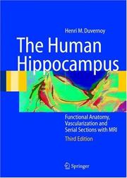
This book offers a precise description of the anatomy of human hippocampus in view of neurosurgical progress and the wealth of medical imaging methods available. A survey of the current concepts explains the functions of the hippocampus and describes its external and internal vascularisation. Head sections and magnetic resonance images complete this comprehensive view of human hippocampal anatomy. It will be of interest to neuroscientists and, in particular, to neurosurgeons, neuroradiologists and neurologists.
Hippocampus (Brain) --- Blood-vessels --- Magnetic resonance imaging --- Ammon's horn --- Cornu ammonis --- Cerebral cortex --- Limbic system --- Neurosurgery. --- Radiology, Medical. --- Neurology. --- Human anatomy. --- Neurosciences. --- Neuroradiology. --- Imaging / Radiology. --- Anatomy. --- Medicine --- Nervous system --- Neuropsychiatry --- Clinical radiology --- Radiology, Medical --- Radiology (Medicine) --- Medical physics --- Nerves --- Neurosurgery --- Neural sciences --- Neurological sciences --- Neuroscience --- Medical sciences --- Anatomy, Human --- Anatomy --- Human biology --- Human body --- Diseases --- Surgery --- Radiology. --- Neurology . --- Radiological physics --- Physics --- Radiation --- Neuroradiography --- Neuroradiology
Book
ISBN: 9783211739716 9783211739709 321173970X 9786612333569 1282333569 3211739718 Year: 2009 Publisher: Wien ; New York : Springer,
Abstract | Keywords | Export | Availability | Bookmark
 Loading...
Choose an application
- Reference Manager
- EndNote
- RefWorks (Direct export to RefWorks)
Advanced MRI requires advanced knowledge of anatomy. This volume correlates thin-section brain anatomy with corresponding clinical 3 T MR images in axial, coronal and sagittal planes to demonstrate the anatomic bases for advanced MR imaging. It specifically correlates advanced neuromelanin imaging, susceptibility-weighted imaging, and diffusion tensor tractography with clinical 3 and 4 T MRI to illustrate the precise nuclear and fiber tract anatomy imaged by these techniques. Each region of the brain stem is then analyzed with 9.4 T MRI to show the anatomy of the medulla, pons, midbrain, and portions of the diencephalonin with an in-plane resolution comparable to myelin- and Nissl-stained light microscopy (40-60 microns). The volume is carefully organized as a teaching text, using concise drawings and beautiful anatomic/MRI images to present the information in sequentially finer detail, so the reader easily assimilates the relationships among the structures shown by high-field MRI.
Neuropathology --- medische beeldvorming --- Psychiatry --- neurologie --- psychiatrie --- Surgery --- neurochirurgie --- Radiotherapy. Isotope therapy --- radiologie --- hersenen --- Physical methods for diagnosis --- Brain Stem --- Cerebellum --- Brain stem --- Tronc cérébral --- Cervelet --- Atlases. --- Atlases --- Atlas --- EPUB-LIV-FT LIVMEDEC SPRINGER-B --- Magnetic Resonance Imaging --- Microscopie. --- Microscopy --- Technique d'imagerie par résonance magnétique nucléaire. --- Tronc cérébral --- Anatomy & histology --- Physiology --- Anatomie et histologie. --- Atlas. --- Methods --- Radiology, Medical. --- Neurology. --- Neurosurgery. --- Psychiatry. --- Neurosciences. --- Neuroradiology. --- Imaging / Radiology. --- Neural sciences --- Neurological sciences --- Neuroscience --- Medical sciences --- Nervous system --- Medicine and psychology --- Mental health --- Psychology, Pathological --- Nerves --- Neurosurgery --- Medicine --- Neuropsychiatry --- Clinical radiology --- Radiology, Medical --- Radiology (Medicine) --- Medical physics --- Diseases --- Radiology. --- Neurology . --- Radiological physics --- Physics --- Radiation --- Neuroradiography --- Neuroradiology
| Listing 1 - 2 of 2 |
Sort by
|

 Search
 Feedback
 About UniCat
  Help
Help News
News