| Listing 1 - 10 of 13 | << page >> |
Sort by
|

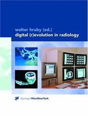
ISBN: 3211834109 Year: 2001 Publisher: Wien : Springer,
Abstract | Keywords | Export | Availability | Bookmark
 Loading...
Choose an application
- Reference Manager
- EndNote
- RefWorks (Direct export to RefWorks)
Radiographic image enhancement. --- Radiography, medical --- Digital techniques.

ISBN: 0849301505 Year: 1990 Publisher: Boca Raton, Fla. : CRC Press,
Abstract | Keywords | Export | Availability | Bookmark
 Loading...
Choose an application
- Reference Manager
- EndNote
- RefWorks (Direct export to RefWorks)
Radiographic Image Enhancement. --- Technology, Radiologic. --- Diagnostic imaging --- Image reconstruction. --- Reconstruction d'image --- Digital techniques --- Mathematics.
Book
ISBN: 3642140211 364214022X Year: 2010 Publisher: Berlin ; Heidelberg : Springer-Verlag,
Abstract | Keywords | Export | Availability | Bookmark
 Loading...
Choose an application
- Reference Manager
- EndNote
- RefWorks (Direct export to RefWorks)
Computed tomography of the heart has become a highly accurate diagnostic modality that is attracting increasing attention. This extensively illustrated book aims to assist the reader in integrating cardiac CT into daily clinical practice, while also reviewing its current technical status and applications. Clear guidance is provided on the performance and interpretation of imaging using the latest technology, which offers greater coverage, better spatial resolution, and faster imaging. The specific features of scanners from all four main vendors, including those that have only recently become available, are presented. Among the wide range of applications and issues to be discussed are coronary artery bypass grafts, stents, plaques, and anomalies, cardiac valves, congenital and acquired heart disease, and radiation exposure. Upcoming clinical uses of cardiac CT, such as plaque imaging and functional assessment, are also explored.
Heart -- Tomography. --- Heart -- Diseases -- Diagnosis. --- Heart -- Imaging. --- Radiographic Image Enhancement --- Tomography, X-Ray --- Diseases --- Diagnostic Techniques and Procedures --- Image Interpretation, Computer-Assisted --- Equipment and Supplies --- Tomography Scanners, X-Ray Computed --- Tomography, X-Ray Computed --- Diagnostic Techniques, Cardiovascular --- Cardiovascular Diseases --- Radiography --- Analytical, Diagnostic and Therapeutic Techniques and Equipment --- Tomography --- Image Enhancement --- Diagnosis --- Diagnostic Imaging --- Photography --- Medicine --- Radiology, MRI, Ultrasonography & Medical Physics --- Health & Biological Sciences --- Cardiology. --- Cardiovascular system --- Diagnosis. --- Medicine. --- Radiology. --- Internal medicine. --- Medicine & Public Health. --- Imaging / Radiology. --- Internal Medicine. --- Heart --- Internal medicine --- Radiology, Medical. --- Medicine, Internal --- Clinical radiology --- Radiology, Medical --- Radiology (Medicine) --- Medical physics --- Radiological physics --- Physics --- Radiation
Periodical
ISSN: 08971889 1618727X Publisher: Philadelphia, Pa
Abstract | Keywords | Export | Availability | Bookmark
 Loading...
Choose an application
- Reference Manager
- EndNote
- RefWorks (Direct export to RefWorks)
Physical methods for diagnosis --- Computer Systems --- Radiographic Image Enhancement --- Radiology Information Systems --- Medical radiology --- Diagnosis, Radioscopic --- Image processing --- Radiologie médicale --- Radiodiagnostics --- Traitement d'images --- Data processing --- Periodicals --- Periodicals. --- Digital techniques --- Informatique --- Périodiques --- Techniques numériques --- Computer Systems. --- Radiographic Image Enhancement. --- Radiology Information Systems. --- Data processing. --- Digital techniques. --- Engineering --- Health Sciences --- Telecommunications Technology --- Biomedical Engineering --- Electronics --- Clinical Medicine --- Diagnostics --- Radiology --- Image Processing & Television Technology --- Radiology, Medical --- Digital image processing --- Digital electronics --- Diagnosis, Radiographic --- Radiodiagnosis --- Radioscopic diagnosis --- Roentgenology, Diagnostic --- X-ray diagnosis --- Diagnosis --- Radiography, Medical --- Clinical radiology --- Radiology (Medicine) --- Medical physics --- Archiving, Radiologic Picture --- Information System, Radiologic --- Information System, Radiology --- Information Systems, Radiologic --- Information Systems, Radiology --- Radiologic Information System --- Radiologic Information Systems --- Radiologic Picture Archiving --- Radiology Information System --- System, Radiologic Information --- System, Radiology Information --- Systems, Radiologic Information --- Systems, Radiology Information --- Xray Information Systems --- PACS (Radiology) --- Picture Archiving and Communication Systems --- Picture Archiving, Radiologic --- X-Ray Information Systems --- Information System, X-Ray --- Information System, Xray --- Information Systems, X-Ray --- Information Systems, Xray --- PACSs (Radiology) --- System, X-Ray Information --- System, Xray Information --- Systems, X-Ray Information --- Systems, Xray Information --- X Ray Information Systems --- X-Ray Information System --- Xray Information System --- Organization, Computer Systems --- Computer Architecture --- Computer Systems Development --- Computer Systems Evaluation --- Computer Systems Organization --- Real-Time Systems --- Architecture, Computer --- Architectures, Computer --- Computer Architectures --- Computer System --- Computer Systems Evaluations --- Development, Computer Systems --- Evaluation, Computer Systems --- Evaluations, Computer Systems --- Real Time Systems --- Real-Time System --- System, Computer --- System, Real-Time --- Systems, Computer --- Systems, Real-Time --- Image Enhancement, Radiographic --- Radiography, Digital --- Digital Radiography --- Enhancement, Radiographic Image --- Enhancements, Radiographic Image --- Image Enhancements, Radiographic --- Radiographic Image Enhancements --- Image Enhancement --- Tomography Scanners, X-Ray Computed --- Picture Archiving And Communication System --- Radiographic Image Enhancement/ --- Radiologia mèdica. --- Processament d'imatges. --- Imatges mèdiques. --- Imatges en medicina --- Sistemes d'imatges en medicina --- Tècniques d'imatge en medicina --- Aparells i instruments mèdics --- Sistemes d'imatges --- Diagnòstic per la imatge --- Fotografia mèdica --- Radiografia mèdica --- Anàlisi d'imatges --- Processament de dades fotogràfiques --- Processament de la imatge --- Tractament d'imatges --- Tractament de la imatge --- Processament òptic de dades --- Processament digital d'imatges --- Visió per ordinador --- Lectors òptics --- Radiologia clínica --- Radiologia (Medicina) --- Física mèdica --- Medicina nuclear --- Radiofàrmacs --- Radiologia dental --- Radiologia intervencionista --- Radiologia pediàtrica --- Radiologia veterinària --- Radioteràpia --- Diagnòstic radiològic
Book
ISBN: 3540799419 9786613087522 3540799427 1283087529 Year: 2011 Publisher: New York : Springer,
Abstract | Keywords | Export | Availability | Bookmark
 Loading...
Choose an application
- Reference Manager
- EndNote
- RefWorks (Direct export to RefWorks)
Standard radiography of the chest remains one of the most widely used imaging modalities but it can be difficult to interpret. The possibility of producing cross-sectional, reformatted 2D and 3D images with CT makes this technique an ideal tool for reinterpreting standard radiography of the chest. The aim of this book is to provide a comprehensive overview of chest radiography interpretation by means of a side-by-side comparison between chest radiographs and CT images. Introductory chapters address the indications for and difficulties of chest radiography as well as the technical and practical aspects of CT reconstruction and image comparison. Thereafter, the radiographic and CT presentations of both anatomical variants and the diseased chest are illustrated and discussed by renowned experts in thoracic imaging. Individual chapters are devoted to the imaging features of selected common diseases and disorders, including COPD, lung cancer, pulmonary embolism and hypertension, atelectasis and chest trauma. The book is complemented by online extra material which provides many further educational examples. .
Chest -- Radiography. --- Radiography, Thoracic. --- Chest --- Tomography, X-Ray --- Image Interpretation, Computer-Assisted --- Radiographic Image Enhancement --- Diagnostic Imaging --- Diseases --- Image Enhancement --- Diagnostic Techniques and Procedures --- Tomography --- Diagnosis --- Photography --- Tomography, X-Ray Computed --- Radiography --- Respiratory Tract Diseases --- Radiography, Thoracic --- Analytical, Diagnostic and Therapeutic Techniques and Equipment --- Medicine --- Health & Biological Sciences --- Radiology, MRI, Ultrasonography & Medical Physics --- Diseases by Body Region --- Diagnostic imaging. --- Radiography. --- Tomography. --- Body section radiography --- Computed tomography --- Computerized tomography --- CT (Computer tomography) --- Laminagraphy --- Laminography --- Radiological stratigraphy --- Stratigraphy, Radiological --- Tomographic imaging --- Zonography --- Skiagraphy --- X-ray photography --- Clinical imaging --- Imaging, Diagnostic --- Medical diagnostic imaging --- Medical imaging --- Noninvasive medical imaging --- Medicine. --- Radiology. --- Medicine & Public Health. --- Imaging / Radiology. --- Cross-sectional imaging --- Radiography, Medical --- Geometric tomography --- Radiology --- X-rays --- Diagnosis, Noninvasive --- Imaging systems in medicine --- Scientific applications --- Radiology, Medical. --- Clinical radiology --- Radiology, Medical --- Radiology (Medicine) --- Medical physics --- Radiological physics --- Physics --- Radiation
Book
ISBN: 3642157254 9786613367921 1283367920 3642157262 Year: 2011 Publisher: Berlin, Heidelberg : Springer Berlin Heidelberg : Imprint: Springer,
Abstract | Keywords | Export | Availability | Bookmark
 Loading...
Choose an application
- Reference Manager
- EndNote
- RefWorks (Direct export to RefWorks)
The field of nuclear medicine has evolved rapidly in recent years, and one very important aspect of this progress has been the introduction of hybrid imaging systems. PET-CT has already gained widespread acceptance in many clinical settings, especially within oncology, and now SPECT-CT promises to emulate its success. Useful applications of this new approach have been identified not only in oncology but also in endocrinology, cardiology, internal medicine, and other specialties. This atlas, which includes hundreds of high-quality images, is a user-friendly guide to the optimal use and interpretation of SPECT-CT. The full range of potential SPECT-CT applications in clinical routine is considered and assessed by acknowledged experts. The book is designed to serve as a reference text for both nuclear physicians and radiologists; it will also provide fundamental support for radiographers, technologists, and nuclear medicine and radiology residents. Following the editors’ other atlases on PET-CT and non-FDG PET-CT, the Atlas of SPECT-CT completes a trilogy offering up-to-date information and guidance on the full spectrum of hybrid imaging.
Oncology. --- Single-photon emission computed tomography -- Atlases. --- Tomography. --- Single-photon emission computed tomography --- Tomography, Emission-Computed --- Investigative Techniques --- Image Interpretation, Computer-Assisted --- Publication Formats --- Radiographic Image Enhancement --- Tomography, X-Ray --- Radiography --- Tomography --- Image Enhancement --- Radionuclide Imaging --- Publication Characteristics --- Analytical, Diagnostic and Therapeutic Techniques and Equipment --- Diagnostic Imaging --- Diagnostic Techniques, Radioisotope --- Photography --- Diagnostic Techniques and Procedures --- Diagnosis --- Tomography, Emission-Computed, Single-Photon --- Tomography, X-Ray Computed --- Methods --- Atlases --- Medicine --- Health & Biological Sciences --- Radiology, MRI, Ultrasonography & Medical Physics --- Single-photon emission computed tomography. --- Radiography, Medical. --- Medical radiography --- Body section radiography --- Computed tomography --- Computerized tomography --- CT (Computer tomography) --- Laminagraphy --- Laminography --- Radiological stratigraphy --- Stratigraphy, Radiological --- Tomographic imaging --- Zonography --- SPECT (Tomography) --- Medicine. --- Radiology. --- Nuclear medicine. --- Medicine & Public Health. --- Nuclear Medicine. --- Imaging / Radiology. --- Cross-sectional imaging --- Radiography, Medical --- Geometric tomography --- Tomography, Emission --- Diagnostic imaging --- Medical photography --- Medical radiology --- Radiology, Medical. --- Oncology . --- Tumors --- Clinical radiology --- Radiology, Medical --- Radiology (Medicine) --- Medical physics --- Atomic medicine --- Radioisotopes in medicine --- Radioactive tracers --- Radioactivity --- Physiological effect --- Computer tomography --- CT (Computed tomography) --- Radiological physics --- Physics --- Radiation
Book
ISBN: 3642017398 9786613087591 3642017401 1283087596 Year: 2011 Publisher: New York : Springer,
Abstract | Keywords | Export | Availability | Bookmark
 Loading...
Choose an application
- Reference Manager
- EndNote
- RefWorks (Direct export to RefWorks)
Dual-energy CT is a novel, rapidly emerging imaging technique which offers important new functional and specific information. With implementation of the technology in commercially available scanners, many clinical applications are now feasible. In this book, physicists and specialists from different CT manufacturers provide an insight into the technological basis of, and the different approaches to, dual-energy CT. Renowned medical scientists in the field explain the pathophysiological and molecular background of the technique, discuss its applications, provide detailed advice on how to obtain optimal results, and offer hints regarding clinical interpretation. The main focus is on the use of dual-energy CT in daily clinical practice, and individual sections are devoted to imaging of the vascular system, the thorax, the abdomen, and the extremities. Evaluations and recommendations are based on personal experience and peer-reviewed literature. Plenty of carefully chosen high-quality images are included to illustrate the clinical benefits of the technique.
Angiography. --- Cardiology. --- Dual-Source-Computertomographie. --- Internal medicine. --- Radiology, Medical. --- Tomography, X-Ray Computed -- Trends. --- Tomography. --- Dual energy CT (Tomography) --- Tomography, X-Ray --- Radiographic Image Enhancement --- Image Interpretation, Computer-Assisted --- Diagnostic Imaging --- Radiography --- Image Enhancement --- Tomography --- Photography --- Diagnostic Techniques and Procedures --- Diagnosis --- Analytical, Diagnostic and Therapeutic Techniques and Equipment --- Tomography, X-Ray Computed --- Medicine --- Health & Biological Sciences --- Radiology, MRI, Ultrasonography & Medical Physics --- Body section radiography --- Computed tomography --- Computerized tomography --- CT (Computer tomography) --- Laminagraphy --- Laminography --- Radiological stratigraphy --- Stratigraphy, Radiological --- Tomographic imaging --- Zonography --- Medicine. --- Radiology. --- Angiology. --- Medicine & Public Health. --- Imaging / Radiology. --- Diagnostic Radiology. --- Internal Medicine. --- Cross-sectional imaging --- Radiography, Medical --- Geometric tomography --- Blood-vessels --- Diagnosis, Radioscopic --- Heart --- Internal medicine --- Medicine, Internal --- Clinical radiology --- Radiology, Medical --- Radiology (Medicine) --- Medical physics --- Diseases --- Radiological physics --- Physics --- Radiation --- Diseases. --- Angiology --- Vascular diseases
Book
ISBN: 3642184561 364218457X Year: 2011 Publisher: Heidelberg : Springer,
Abstract | Keywords | Export | Availability | Bookmark
 Loading...
Choose an application
- Reference Manager
- EndNote
- RefWorks (Direct export to RefWorks)
This book, unique in focusing specifically on cardiac masses, is the result of cooperation among a number of teams of radiologists working under the aegis of the French Society of Cardiovascular Imaging (SFICV). Its goal is to 1) review the different CMR sequences and CT acquisition protocols used to explore cardiac masses, 2) to demonstrate the several CMR and MDCT features of cardiac masses. It has been designed as a teaching tool and offers a fully illustrated compendium of clinical cases, tables summarizing data, and decision-making trees essential in everyday practice. It is presented as a practical handbook and can be either read cover to cover or consulted whenever needed during a cardiac imaging assignment. The book is intended for all students and experienced practitioners, whether radiologists or not, who are interested in cardiac or thoracic pathology.
Echocardiography -- Examinations, questions, etc. --- Echocardiography. --- Medical physics. --- Progetto CMR (Firm). --- Tomography. --- Diagnostic Imaging --- Thoracic Neoplasms --- Heart Diseases --- Tomography, X-Ray --- Radiographic Image Enhancement --- Tomography --- Image Interpretation, Computer-Assisted --- Image Enhancement --- Neoplasms by Site --- Radiography --- Cardiovascular Diseases --- Diagnostic Techniques and Procedures --- Diagnosis --- Diseases --- Neoplasms --- Photography --- Analytical, Diagnostic and Therapeutic Techniques and Equipment --- Heart Neoplasms --- Tomography, X-Ray Computed --- Magnetic Resonance Imaging --- Medicine --- Health & Biological Sciences --- Radiology, MRI, Ultrasonography & Medical Physics --- Heart --- Imaging. --- Abnormalities --- Diagnosis. --- Cardiac diagnostic imaging --- Cardiac imaging --- Diagnostic cardiac imaging --- Imaging of the heart --- Imaging --- Medicine. --- Radiology. --- Interventional radiology. --- Cardiology. --- Oncology. --- Cardiac surgery. --- Medicine & Public Health. --- Imaging / Radiology. --- Interventional Radiology. --- Cardiac Surgery. --- Radiology, Medical. --- Oncology . --- Surgery. --- Cardiac surgery --- Open-heart surgery --- Tumors --- Internal medicine --- Radiology, Interventional --- Medical radiology --- Therapeutics --- Clinical radiology --- Radiology, Medical --- Radiology (Medicine) --- Medical physics --- Surgery --- Interventional radiology . --- Radiological physics --- Physics --- Radiation
Book
ISBN: 3540892311 9786613367877 1283367874 354089232X Year: 2011 Publisher: Heidelberg : Springer,
Abstract | Keywords | Export | Availability | Bookmark
 Loading...
Choose an application
- Reference Manager
- EndNote
- RefWorks (Direct export to RefWorks)
CT of the Acute Abdomen provides an in-depth and comprehensive account of the use of CT in patients with acute abdomen. Recent significant developments in CT that are of relevance in imaging of the acute abdomen, including multislice CT and multiplanar reconstructions, receive particular attention. CT features are clearly illustrated, and pitfalls and differential diagnoses are discussed. The first section of the book presents epidemiological and clinical data in acute abdomen. The second and third sections document the key CT findings and their significance and discuss the technological background, including acquisition, reformatting, and surface and volume rendering. The fourth and fifth sections, which form the main body of the book, examine in detail the various clinical applications of CT in nontraumatic and traumatic acute abdomen. Full account is taken of new trends in the diagnosis and management of patients with acute abdomen, and the role of CT is examined in this context. This book will serve as an ideal guide to the performance and interpretation of CT in the setting of the acute abdomen; it will be of value to all general and gastrointestinal radiologists, as well as emergency room physicians and gastrointestinal surgeons.
Abdomen, Acute -- Radiography. --- Acute abdomen. --- Digestive system diseases -- Radiography. --- Acute abdomen --- Tomography, X-Ray --- Abdominal Pain --- Image Interpretation, Computer-Assisted --- Investigative Techniques --- Diseases --- Diagnostic Imaging --- Diagnosis --- Radiographic Image Enhancement --- Pain --- Signs and Symptoms, Digestive --- Diagnostic Techniques and Procedures --- Image Enhancement --- Tomography --- Analytical, Diagnostic and Therapeutic Techniques and Equipment --- Signs and Symptoms --- Photography --- Pathological Conditions, Signs and Symptoms --- Tomography, X-Ray Computed --- Abdomen, Acute --- Radiography --- Diagnosis, Differential --- Digestive System Diseases --- Methods --- Medicine --- Health & Biological Sciences --- Diseases by Body Region --- Radiology, MRI, Ultrasonography & Medical Physics --- Abdomen --- Diagnosis. --- Tomography. --- Ultrasonic imaging. --- Surgical abdomen --- Medicine. --- Radiology. --- Internal medicine. --- Gastroenterology. --- Medicine & Public Health. --- Imaging / Radiology. --- Diagnostic Radiology. --- Internal Medicine. --- Viscera --- Abdominal pain --- Surgical diseases --- Surgery --- Radiology, Medical. --- Medicine, Internal --- Internal medicine --- Digestive organs --- Clinical radiology --- Radiology, Medical --- Radiology (Medicine) --- Medical physics --- Gastroenterology . --- Radiological physics --- Physics --- Radiation
Book
ISBN: 1849962596 9786612972171 1282972170 184996260X Year: 2011 Publisher: New York : Springer,
Abstract | Keywords | Export | Availability | Bookmark
 Loading...
Choose an application
- Reference Manager
- EndNote
- RefWorks (Direct export to RefWorks)
Vascular medicine is an ever changing and rapidly advancing field, and with the onset of state of the art diagnostic and imaging modalities, we are recognizing that vascular illness is more prevalent than once thought. The proper diagnosis and treatment of this vascular illness may prevent stroke, salvage a limb and even save a life. Advancements in CT angiography have made vascular CT angiography the modality of choice for accurately diagnosing vascular disease and the management of its treatment. Modern, multi-slice CT provides noninvasive, direct imaging of virtually the entire vascular system in a safe, effective and accurate manner without the inherent risk attributed to invasive angiography. Furthermore, it is widely available, even in institutions that do not perform invasive angiography. Vascular CT Angiography Manual distills vascular CT angiography and the diseases for which it is meant to diagnose into an easy to follow and inclusive resource. Designed for residents and fellows in cardiovascular medicine and radiology as well as for those already in practice, this work makes 'difficult to understand' concepts easy to comprehend with the aid of simple, intuitive diagrams, charts and clinical CT image examples. For those already proficient in vascular CT, this book will serve as a valuable resource as it compiles a complete radiation review, imaging protocols, screening recommendations, disease states and imaging tips in one location. It is organized and easy to follow, as each clinical chapter is organized to include sections on anatomy, disease states, accuracy of CT, and an approach to reading CT angiography.
Angiography. --- Blood-vessels -- Diseases -- Diagnosis. --- Interventional radiology. --- Cardiovascular system --- Angiography --- Blood-vessels --- Tomography --- Publication Formats --- Tomography, X-Ray --- Radiographic Image Enhancement --- Anatomy --- Diseases --- Diagnostic Techniques, Cardiovascular --- Diagnostic Imaging --- Investigative Techniques --- Image Interpretation, Computer-Assisted --- Diagnostic Techniques and Procedures --- Image Enhancement --- Analytical, Diagnostic and Therapeutic Techniques and Equipment --- Publication Characteristics --- Tomography, X-Ray Computed --- Handbooks --- Cardiovascular Diseases --- Cardiovascular System --- Radiography --- Methods --- Diagnosis --- Photography --- Medicine --- Health & Biological Sciences --- Radiography. --- Medicine. --- Radiology. --- Internal medicine. --- Cardiology. --- Cardiac surgery. --- Vascular surgery. --- Medicine & Public Health. --- Imaging / Radiology. --- Diagnostic Radiology. --- Cardiac Surgery. --- Vascular Surgery. --- Internal Medicine. --- Diagnosis, Radioscopic --- Radiography, Medical --- Radiology, Medical. --- Heart --- Surgery. --- Medicine, Internal --- Vascular surgery --- Cardiac surgery --- Open-heart surgery --- Clinical radiology --- Radiology, Medical --- Radiology (Medicine) --- Medical physics --- Internal medicine --- Surgery --- Radiological physics --- Physics --- Radiation
| Listing 1 - 10 of 13 | << page >> |
Sort by
|

 Search
 Feedback
 About UniCat
  Help
Help News
News