| Listing 1 - 10 of 12 | << page >> |
Sort by
|

ISBN: 0585382743 9780585382746 0890964742 9780890964743 0890964750 9780890964750 0890964882 9780890964880 Year: 1991 Publisher: College Station, Tex. Texas A & M University Press
Abstract | Keywords | Export | Availability | Bookmark
 Loading...
Choose an application
- Reference Manager
- EndNote
- RefWorks (Direct export to RefWorks)

ISBN: 0197561977 1280502614 0195303539 0198035691 1433701146 9780198035695 9780195157062 0195157060 9781280502613 9780195303537 9786610502615 6610502617 0190288965 Year: 2004 Publisher: Oxford, [England] : Oxford University Press,
Abstract | Keywords | Export | Availability | Bookmark
 Loading...
Choose an application
- Reference Manager
- EndNote
- RefWorks (Direct export to RefWorks)
This is an in-depth account of the fossil skull anatomy and evolutionary significance of the 3.6-3.0 million year old early human species Australopithecus afarensis. Knowledge of this species is pivotal to understanding early human evolution, because 1) the sample of fossil remains of A. afarensis is among the most extensive for any early human species, and the majority of remains are of taxonomically informative skulls and teeth; 2) the wealth of material makes A. afarensis an indispensable point of reference for the interpretation of other fossil discoveries; 3) the species occupies a time period that is the focus of current research to determine when, where, and why the human lineage first diversified into separate contemporaneous lines of descent.
Australopithecus afarensis. --- Skull. --- Craniology. --- Physical anthropology --- Brain case --- Braincase --- Brainpan --- Cranium --- Fontanel --- Fontanelle --- Bones --- Skeleton --- Head --- Australopithecines --- Fossil hominids --- Australopithecus afarensis --- Crâne --- Craniologie --- Australopithecus afarensis., --- lemac

ISBN: 9780702036675 0702036676 9780443074998 0443074992 Year: 2004 Publisher: [Place of publication not identified] Elsevier Churchill Livingstone
Abstract | Keywords | Export | Availability | Bookmark
 Loading...
Choose an application
- Reference Manager
- EndNote
- RefWorks (Direct export to RefWorks)
Osteopathic medicine --- Skull --- Manipulation, Osteopathic --- Head. --- Sacrum. --- Skull. --- Brain case --- Braincase --- Brainpan --- Cranium --- Fontanel --- Fontanelle --- Bones --- Skeleton --- Head --- Osteopathy --- Medicine --- Calvarium --- Calvaria --- Skulls --- Sacra --- Sacral Vertebra --- Sacral Vertebrae --- Sacrums --- Vertebra, Sacral --- Vertebrae, Sacral --- Heads --- methods. --- Sacrum --- Methods
Book
ISBN: 3319518909 3319518887 Year: 2017 Publisher: Cham : Springer International Publishing : Imprint: Springer,
Abstract | Keywords | Export | Availability | Bookmark
 Loading...
Choose an application
- Reference Manager
- EndNote
- RefWorks (Direct export to RefWorks)
This astute volume brings together the latest expert research on adamantinomatous craniopharyngiomas (ACPs). ACPs are histologically benign but clinically aggressive tumors exhibiting a high propensity for local invasion into the hypothalamus, optic and vascular structures. These tumors, as well as the current treatments, may result in pan-hypopituitarism, diabetes insipidus, morbid obesity followed by type II diabetes mellitus, blindness, as well as serious behavioral and psychosocial impairments. Exploring in detail advances in both the understanding of tumor biology as well as clinical advances in patient management are explored in detail, this book will also look towards potential new treatment approaches. Basic Research and Clinical Aspects of Adamantinomatous Craniopharyngioma is the first book compiling all current research on ACPs. Mouse and human studies have unequivocally demonstrated that mutations in CTNNB1 encoding -catenin underlie the etiology of the majority, if not all ACP tumors. Genetic studies in mice have shown that ACPs are tumors of the pituitary gland and not of the hypothalamus as previously thought, and are derived from Rathke’s pouch precursors. In addition, a role for tissue-specific adult pituitary stem cells has been revealed as causative of ACP. Together, these studies have provided novel insights into the molecular and cellular etiology as well as the pathogenesis of human ACP. Finally, this volume covers new treatment approaches that have been shown to be effective both in reducing ACP burden as well as reducing the morbidity associated with therapy. .
Brain --- Skull --- Cancer in children. --- Tumors. --- Childhood cancer --- Pediatric cancer --- Brain case --- Braincase --- Brainpan --- Cranium --- Fontanel --- Fontanelle --- Medicine. --- Cancer research. --- Endocrinology. --- Stem cells. --- Medicine & Public Health. --- Cancer Research. --- Stem Cells. --- Tumors in children --- Bones --- Skeleton --- Head --- Oncology. --- Tumors --- Internal medicine --- Hormones --- Colony-forming units (Cells) --- Mother cells --- Progenitor cells --- Cells --- Endocrinology . --- Cancer research

ISBN: 1281351059 9786611351052 3540494685 3540341900 Year: 2007 Publisher: Berlin ; New York : Springer,
Abstract | Keywords | Export | Availability | Bookmark
 Loading...
Choose an application
- Reference Manager
- EndNote
- RefWorks (Direct export to RefWorks)
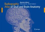
The English Edition contains a few differences from the first ItaHan Edition, which require an explanation. Firstly, some imag es, especially some 3D reconstructions, have been modified in order to make them clearer. Secondly, in agreement with the Publisher, we have disowned one of our statements in the preface to the Italian Edition. Namely, we have now added a brief introductory text for each section, by way of explanation to the anatomical and physiological notes. This should make it easier for the reader to understand and refer to this Atlas. These differences derive from our experience with the previous edition and are meant to be an improvement thereof Hopefully, there will be more editions to follow, so that we may further improve our work and keep ourselves busy on lone some evenings. Finally, the improvements in this edition are a reminder to the reader that one should never purchase the first edition of a work. UAquila, January 2006 The Authors Preface to the Italian Edition I have been meaning to publish an atlas of neuroradiologic cranio-encephaHc anatomy for at least the last decade. Normal anatomy has always been of great and charming interest to me. Over the years, while preparing lectures for my students, I have always enjoyed lingering on anatomical details that today are rendered with astonishing realism by routine diagnostic ima ging.
Skull --- Brain --- Anatomy --- Radiography --- Cerebrum --- Mind --- Central nervous system --- Head --- Brain case --- Braincase --- Brainpan --- Cranium --- Fontanel --- Fontanelle --- Bones --- Skeleton --- Radiology, Medical. --- Nuclear medicine. --- Radiotherapy. --- Neurology. --- Neurosurgery. --- Neuroradiology. --- Imaging / Radiology. --- Nuclear Medicine. --- Nerves --- Neurosurgery --- Medicine --- Nervous system --- Neuropsychiatry --- Radiation therapy --- Electrotherapeutics --- Hospitals --- Medical electronics --- Medical radiology --- Therapeutics, Physiological --- Phototherapy --- Atomic medicine --- Radioisotopes in medicine --- Radioactive tracers --- Radioactivity --- Clinical radiology --- Radiology, Medical --- Radiology (Medicine) --- Medical physics --- Surgery --- Diseases --- Radiological services --- Physiological effect --- Radiology. --- Neurology . --- Radiological physics --- Physics --- Radiation --- Neuroradiography --- Neuroradiology
Book
ISBN: 9783319082424 3319082418 9783319082417 3319082426 Year: 2015 Publisher: Cham : Springer International Publishing : Imprint: Springer,
Abstract | Keywords | Export | Availability | Bookmark
 Loading...
Choose an application
- Reference Manager
- EndNote
- RefWorks (Direct export to RefWorks)
This book, featuring more than 180 high spatial resolution images obtained with state-of-the-art MDCT and MRI scanners, depicts in superb detail the anatomy of the temporal bone, recognized to be one of the most complex anatomic areas. In order to facilitate identification of individual anatomic structures, the images are presented in the same way in which they emanate from contemporary imaging modalities, namely as consecutive submillimeter sections in standardized slice orientations, with all anatomic landmarks labeled. While various previous publications have addressed the topic of temporal bone anatomy, none has presented complete isotropic submillimeter 3D volume datasets of MDCT or MRI examinations. The Temporal Bone MDCT and MRI Anatomy offers radiologists, head and neck surgeons, neurosurgeons, and anatomists a comprehensive guide to temporal bone sectional anatomy that resembles as closely as possible the way in which it is now routinely reviewed, i.e., on the screens of diagnostic workstations or picture archiving and communication systems (PACS).
Medicine & Public Health. --- Imaging / Radiology. --- Head and Neck Surgery. --- Neurosurgery. --- Anatomy. --- Medicine. --- Human anatomy. --- Radiology, Medical. --- Head --- Médecine --- Anatomie humaine --- Tête --- Surgery. --- Chirurgie --- Skull --- Magnetic resonance imaging. --- Tomography. --- Brain case --- Braincase --- Brainpan --- Cranium --- Fontanel --- Fontanelle --- Radiology. --- Otolaryngologic surgery. --- Bones --- Skeleton --- Anatomy, Human --- Anatomy --- Human biology --- Medical sciences --- Human body --- Nerves --- Neurosurgery --- Clinical radiology --- Radiology, Medical --- Radiology (Medicine) --- Medical physics --- Surgery --- Radiological physics --- Physics --- Radiation --- Operative otolaryngology --- Otolaryngologic surgery --- Surgery, Operative --- Surgery, Orificial
Book
ISBN: 3030007227 3030007219 Year: 2019 Publisher: Cham : Springer International Publishing : Imprint: Springer,
Abstract | Keywords | Export | Availability | Bookmark
 Loading...
Choose an application
- Reference Manager
- EndNote
- RefWorks (Direct export to RefWorks)
This book is designed to serve as an up-to-date reference on the use of cone-beam computed tomography for the purpose of 3D imaging of the craniofacial complex. The focus is in particular on the ways in which craniofacial 3D imaging changes how we think about conventional diagnosis and treatment planning and on its clinical applications within orthodontics and oral and maxillofacial surgery. Emphasis is placed on the value of 3D imaging in visualizing the limits of the alveolar bone, the airways, and the temporomandibular joints and the consequences for treatment planning and execution. The book will equip readers with the knowledge required in order to apply and interpret 3D imaging to the benefit of patients. All of the authors have been carefully selected on the basis of their expertise in the field. In describing current thinking on the merits of 3D craniofacial imaging, they draw both on the available scientific literature and on their own translational research findings.
Surgery. --- Dentistry. --- Radiology, Medical. --- Oral and Maxillofacial Surgery. --- Diagnostic Radiology. --- Skull --- Face --- Three-dimensional imaging in medicine. --- Imaging. --- Three-dimensional medical imaging --- Imaging systems in medicine --- Human face --- Head --- Pathognomy --- Physiognomy --- Brain case --- Braincase --- Brainpan --- Cranium --- Fontanel --- Fontanelle --- Bones --- Skeleton --- Clinical radiology --- Radiology, Medical --- Radiology (Medicine) --- Medical physics --- Dental surgery --- Odontology --- Surgery, Dental --- Medicine --- Oral medicine --- Teeth --- Surgery, Primitive --- Oral surgery. --- Maxillofacial surgery. --- Radiology. --- Radiological physics --- Physics --- Radiation --- Oral surgery --- Surgery, Oral --- Oral surgeons
Book
ISBN: 1282894692 9786612894695 0226233499 9780226233499 9781282894693 9780226233482 0226233480 Year: 2010 Publisher: Chicago The University of Chicago Press
Abstract | Keywords | Export | Availability | Bookmark
 Loading...
Choose an application
- Reference Manager
- EndNote
- RefWorks (Direct export to RefWorks)
When Philadelphia naturalist Samuel George Morton died in 1851, no one cut off his head, boiled away its flesh, and added his grinning skull to a collection of crania. It would have been strange, but perhaps fitting, had Morton's skull wound up in a collector's cabinet, for Morton himself had collected hundreds of skulls over the course of a long career. Friends, diplomats, doctors, soldiers, and fellow naturalists sent him skulls they gathered from battlefields and burial grounds across America and around the world. With The Skull Collectors, eminent historian Ann Fabian resurrects that popular and scientific movement, telling the strange-and at times gruesome-story of Morton, his contemporaries, and their search for a scientific foundation for racial difference. From cranial measurements and museum shelves to heads on stakes, bloody battlefields, and the "rascally pleasure" of grave robbing, Fabian paints a lively picture of scientific inquiry in service of an agenda of racial superiority, and of a society coming to grips with both the deadly implications of manifest destiny and the mass slaughter of the Civil War. Even as she vividly recreates the past, Fabian also deftly traces the continuing implications of this history, from lingering traces of scientific racism to debates over the return of the remains of Native Americans that are held by museums to this day. Full of anecdotes, oddities, and insights, The Skull Collectors takes readers on a darkly fascinating trip down a little-visited but surprisingly important byway of American history.
Craniology --- Anthropometry --- Skull --- Brain case --- Braincase --- Brainpan --- Cranium --- Fontanel --- Fontanelle --- Bones --- Skeleton --- Head --- Skeletal remains --- Physical anthropology --- Body size --- Social aspects --- History --- Catalogs and collections --- Morton, Samuel George, --- american studies, history, historical, skulls, bones, race, science, naturalist, samuel george morton, racial creations, polygenism, 19th century, natural scientist, difference, manifest destiny, civil war, native americans, indigenous peoples, racialized sciences, craniology, collections, crania americana, comparative, scientific racism, lithography, images, illustrations, inferiority, separation.
Book
ISBN: 0387765859 0387765840 1489991816 Year: 2008 Publisher: New York : Springer,
Abstract | Keywords | Export | Availability | Bookmark
 Loading...
Choose an application
- Reference Manager
- EndNote
- RefWorks (Direct export to RefWorks)
Primates have unusual heads among mammals. Their big brains, relatively short faces and forward-facing eyes are part of a unique combination of traits that have captured the interest of biological anthropologists for decades. Describing the patterns of primate craniofacial evolution as well as sorting out the functional consequences of this evolutionary history has been fundamental in developing our current understanding of primates. Primate Craniofacial Function and Biology surveys current research on primate heads emphasizing the recent progress and diversity of functional studies into primate and mammalian craniofacial form. Much of the work included in this volume was inspired by William L. Hylander and his life-long contribution to research on primate craniofacial form and function.
Masticatory muscles. --- Physical anthropology. --- Primates --- Skull. --- Anatomy. --- Evolution. --- Biological anthropology --- Somatology --- Anthropology --- Human biology --- Craniomandibular muscles --- Masticatory apparatus --- Muscles of mastication --- Muscles --- Brain case --- Braincase --- Brainpan --- Cranium --- Fontanel --- Fontanelle --- Bones --- Skeleton --- Head --- Evolution (Biology). --- Zoology. --- Animal physiology. --- Morphology (Animals). --- Developmental biology. --- Anthropology. --- Evolutionary Biology. --- Animal Physiology. --- Animal Anatomy / Morphology / Histology. --- Developmental Biology. --- Human beings --- Development (Biology) --- Biology --- Growth --- Ontogeny --- Animal morphology --- Animals --- Body form in animals --- Zoology --- Morphology --- Animal physiology --- Anatomy --- Natural history --- Animal evolution --- Biological evolution --- Darwinism --- Evolutionary biology --- Evolutionary science --- Origin of species --- Evolution --- Biological fitness --- Homoplasy --- Natural selection --- Phylogeny --- Physiology --- Evolutionary biology. --- Animal anatomy. --- Animal anatomy
Periodical
ISSN: 22124276 22124268
Abstract | Keywords | Export | Availability | Bookmark
 Loading...
Choose an application
- Reference Manager
- EndNote
- RefWorks (Direct export to RefWorks)
Dentistry --- Mouth --- Face --- Skull --- Oral Medicine. --- Mouth. --- Craniofacial Abnormalities. --- Dentistry. --- Face. --- Skull. --- abnormalities. --- Abnormalities, Craniofacial --- Abnormality, Craniofacial --- Craniofacial Abnormality --- Cavitas Oris --- Cavitas oris propria --- Mouth Cavity Proper --- Oral Cavity Proper --- Vestibule Oris --- Vestibule of the Mouth --- Oral Cavity --- Cavity, Oral --- Medicine, Oral --- Stomatology --- Brain case --- Braincase --- Brainpan --- Cranium --- Fontanel --- Fontanelle --- Human face --- Dental surgery --- Odontology --- Surgery, Dental --- Bones --- Skeleton --- Head --- Pathognomy --- Physiognomy --- Medicine --- Oral medicine --- Teeth --- Health Sciences --- Cirurgia maxil·lofacial. --- Cirurgia oral. --- Cirurgia bucal --- Cirurgia maxil·lofacial --- Cirurgia dental --- Cirurgia de la cara --- Cirurgia oral
| Listing 1 - 10 of 12 | << page >> |
Sort by
|

 Search
 Feedback
 About UniCat
  Help
Help News
News