| Listing 1 - 2 of 2 |
Sort by
|
Book
ISBN: 9780521195997 9781139018173 9781139839600 1139839608 1139018175 9781139841986 113984198X 9781139844345 1139844342 9781139844345 9781139840798 1139840797 0521195993 1283812517 9781283812511 1107232872 1139853430 1139845497 9781107232877 9781139853439 9781139845496 Year: 2013 Publisher: Cambridge : Cambridge University Press,
Abstract | Keywords | Export | Availability | Bookmark
 Loading...
Choose an application
- Reference Manager
- EndNote
- RefWorks (Direct export to RefWorks)
Recent developments in scanning electron microscopy (SEM) have resulted in a wealth of new applications for cell and molecular biology, as well as related biological disciplines. It is now possible to analyze macromolecular complexes within their three-dimensional cellular microenvironment in near native states at high resolution and to identify specific molecules and their structural and molecular interactions. New approaches include cryo-SEM applications and environmental SEM (ESEM), staining techniques and processing applications combining embedding and resin-extraction for imaging with high resolution SEM, and advances in immuno-labeling. New developments include helium ion microscopy, automated block-face imaging combined with serial sectioning inside an SEM chamber, and Focused Ion Beam Milling (FIB) combined with block-face SEM. With chapters written by experts, this guide gives an overview of SEM and sample processing for SEM and highlights several advances in cell and molecular biology that greatly benefited from using conventional, cryo, immuno and high-resolution SEM.
Biology --- Scanning electron microscopy. --- Methodology. --- Scanning electron microscopy --- methodology --- Electron microscopy --- Biology, Experimental --- Biology - methodology

ISBN: 1281139904 9786611139902 0387729720 0387729704 1489995846 Year: 2008 Publisher: New York : Springer,
Abstract | Keywords | Export | Availability | Bookmark
 Loading...
Choose an application
- Reference Manager
- EndNote
- RefWorks (Direct export to RefWorks)
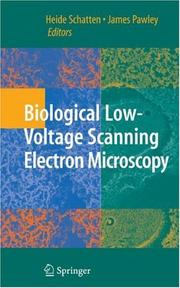
Field-emission, low-voltage scanning electron microscopy (LVSEM) is a field that has grown tremendously in recent years because is offers the optimal method for viewing complex surfaces at high resolution and in three dimensions. However, even though the instrumentation required to get good results at low beam voltage has become increasingly available, there has been a lag in its application to biological specimens. What seemed to be missing was volume that combined both the theory and practice of using this equipment in an optimal manner with a thorough treatment of biological specimen preparation. Biological Low-Voltage Scanning Electron Microscopy is the first book to address both of these aspects of biological LVSEM. After providing a thorough description of the unique advantages and the operating constraints related to operating a scanning electron microscope at low beam voltage, the remainder of book focuses on the the best way to image all types of plant and animal cells and covers specimens that range from macromolecules to the surfaces revealed by de-embedding resin-embedded samples. Advanced specimen preparation techniques such as cryo-LVSEM, and immuno-gold-LVSEM are fully covered, as is x-ray microanalysis at low beam voltage and live-time stereo imaging. The preparative protocols provided represent the distilled essence of the experience of a group of world-renowned authors who have, for many decades, been instrumental in developing and applying new approaches to LVSEM to support their own biological research.
Low-voltage scanning electron microscopy. --- Field emission cathodes. --- Scanning electron microscopes. --- Scanning electron microscope --- Electron microscopes --- Cathodes --- Field emission --- Low voltage systems --- Scanning electron microscopy --- Microscopy. --- Zoology. --- Cytology. --- Microbiology. --- Biological Microscopy. --- Cell Biology. --- Microbial biology --- Biology --- Microorganisms --- Cell biology --- Cellular biology --- Cells --- Cytologists --- Natural history --- Animals --- Analysis, Microscopic --- Light microscopy --- Micrographic analysis --- Microscope and microscopy --- Microscopic analysis --- Optical microscopy --- Optics --- Cell biology.
| Listing 1 - 2 of 2 |
Sort by
|

 Search
 Feedback
 About UniCat
  Help
Help News
News