| Listing 1 - 5 of 5 |
Sort by
|

ISBN: 9781588293039 1588293033 9781592598182 9786610359882 1280359889 1592598188 Year: 2005 Publisher: Totowa, N.J. : Humana Press,
Abstract | Keywords | Export | Availability | Bookmark
 Loading...
Choose an application
- Reference Manager
- EndNote
- RefWorks (Direct export to RefWorks)
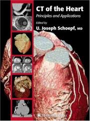
The introduction of fast ECG-synchronized computed tomography (CT) techniques enables imaging of the heart with a combination of speed and spatial resolution unparalleled by other noninvasive imaging modalities. Applying these modalities for the evaluation of coronary artery disease is a topic of active current research. Coronary artery calcium measurements are investigated as a marker for cardiac risk stratification. With contrast-enhanced CT coronary angiography, coronary arteries can be visualized with unprecedented detail, so that noninvasive stenosis assessment appears within reach. With increasing accuracy CT enables evaluation of coronary artery bypass grafts and stents. The cross-sectional nature of CT may to some degree allow noninvasive assessment of the coronary artery wall. CT for evaluating cardiac perfusion, motion, and viability is being investigated. In CT of the Heart, leading radiologists, cardiologists, physicists, engineers, and basic and clinical scientists from around the world survey the full scope of current developments, research, and scientific controversy regarding principles and applications of cardiac CT. Richly illustrated with numerous black-and-white and color images, the book discusses the interpretation of CT of the heart in a variety of clinical, physiologic, and pathologic applications. The authors emphasize current state-of-the-art uses of computed tomography, but also examine emerging developments at the horizon. They review the technical basis of CT image acquisition as well as the tools for image visualization and analysis. Meticulous and comprehensive, CT of the Heart authoritatively defines the current status of computed tomography of the heart, offering a truly balanced view of its technology, applications, significance, and future potential.
Heart --- Tomography, X-Ray Computed. --- Coeur --- radiography. --- Tomography. --- Tomographie --- Heart -- Tomography. --- Diagnostic Imaging --- Radiographic Image Enhancement --- Cardiovascular System --- Tomography, X-Ray --- Image Interpretation, Computer-Assisted --- Diagnostic Techniques and Procedures --- Image Enhancement --- Anatomy --- Tomography --- Photography --- Diagnosis --- Analytical, Diagnostic and Therapeutic Techniques and Equipment --- Tomography, X-Ray Computed --- Radiography --- Medicine --- Health & Biological Sciences --- Cardiovascular Diseases --- Radiology, MRI, Ultrasonography & Medical Physics --- Cardiology. --- Cardiographic tomography --- Diseases --- Medicine. --- Radiology. --- Medicine & Public Health. --- Imaging / Radiology. --- Radiological physics --- Physics --- Radiation --- Clinical sciences --- Medical profession --- Human biology --- Life sciences --- Medical sciences --- Pathology --- Physicians --- Internal medicine --- Radiology, Medical. --- Clinical radiology --- Radiology, Medical --- Radiology (Medicine) --- Medical physics
Book
ISBN: 9781848000926 184800091X 9781848000919 1848000928 Year: 2008 Publisher: London : Springer,
Abstract | Keywords | Export | Availability | Bookmark
 Loading...
Choose an application
- Reference Manager
- EndNote
- RefWorks (Direct export to RefWorks)
Cardiovascular computed tomography (CT) has evolved from novel technology to research tool to essential clinical imaging modality at an astounding pace. The technology has great relevance for many medical and surgical disciplines focused on the treatment of cardiovascular disease. Thanks to the ability of CT to image anatomical, physiological and tissue characteristics of large and small vessel within seconds, and reconstruct multimodal two- and three-dimensional images within minutes, cardiovascular CT has facilitated practical clinical applications important to cardiovascular diagnosis, risk stratification and procedure guidance. Handbook of Cardiovascular CT: Essentials for Clinical Practice has been created as a primer to assist cardiologists, radiologists and other readers involved in cardiac imaging. The new wave in CT technology requires education and training focused on providing an understanding of the essentials of methodology, technique, and clinical image analysis. Reviewing the practical techniques and interpretation of the modality, this book’s compact format provides a handy reference tool and provides the answers to important cardiovascular CT questions. The breadth and depth of the knowledge of the individual authors also ensures that the concise chapters are geared towards covering the essential topics, but with additional depth provided by the inclusion of teaching pearls as well as selected key images.
Medicine & Public Health. --- Cardiology. --- Imaging / Radiology. --- Diagnostic Radiology. --- Internal Medicine. --- Ultrasound. --- Cardiac Surgery. --- Medicine. --- Medical radiology --- Diagnosis, Ultrasonic. --- Internal medicine. --- Heart --- Médecine --- Radiologie médicale --- Médecine interne --- Cardiologie --- Coeur --- Surgery. --- Chirurgie --- Cardiovascular system -- Diseases -- Diagnosis. --- Cardiovascular system -- Diseases. --- Heart -- Diseases. --- Heart -- Tomography. --- Tomography, X-Ray Computed --- Cardiovascular Diseases --- Tomography, X-Ray --- Radiographic Image Enhancement --- Image Interpretation, Computer-Assisted --- Diseases --- Radiography --- Diagnostic Imaging --- Image Enhancement --- Tomography --- Photography --- Diagnostic Techniques and Procedures --- Diagnosis --- Analytical, Diagnostic and Therapeutic Techniques and Equipment --- Medicine --- Health & Biological Sciences --- Cardiovascular system --- Diagnosis. --- Tomography. --- Circulatory system --- Vascular system --- Radiology. --- Cardiac surgery. --- Blood --- Circulation --- Radiology, Medical. --- Cardiac surgery --- Open-heart surgery --- Diagnosis, Ultrasonic --- Diagnostic sonography --- Diagnostic ultrasonics --- Diagnostic ultrasonography --- Diagnostic ultrasound --- Medical diagnostic ultrasonic imaging --- Medical ultrasonography --- Ultrasonic diagnosis --- Ultrasonic diagnostic imaging --- Ultrasonic imaging --- Ultrasonic waves --- Diagnostic imaging --- Ultrasonics in medicine --- Medicine, Internal --- Clinical radiology --- Radiology, Medical --- Radiology (Medicine) --- Medical physics --- Internal medicine --- Surgery --- Diagnostic use --- Radiological physics --- Physics --- Radiation
Book
ISBN: 9788847008328 884700831X 9788847008311 9786612458866 1282458868 8847008328 Year: 2008 Publisher: Milano Springer-Verlag Milan
Abstract | Keywords | Export | Availability | Bookmark
 Loading...
Choose an application
- Reference Manager
- EndNote
- RefWorks (Direct export to RefWorks)
MDCT: From Protocols to Practice tackles contemporary and topical issues in MDCT technology and applications. As an updated edition of MDCT: A Practical Approach, this volume offers new content as well as revised chapters from the previous volume. New chapters discuss important topics such as imaging of children and obese subjects, the use of contrast medium in pregnant women coronary, MDCT angiography, and PET/CT in abdominal and pelvic malignancies. Furthermore an Appendix with over 50 updated MDCT scanning protocols completes this publication. The book emphasizes the practical aspects of MDCT, making it an invaluable source of information for radiologists, residents, medical physicists, and radiology technologists in everyday clinical practice.
Medicine & Public Health. --- Imaging / Radiology. --- Diagnostic Radiology. --- Interventional Radiology. --- Medicine. --- Medical radiology --- Interventional radiology. --- Médecine --- Radiologie médicale --- Radiologie interventionnelle --- Kalra, M. K. (Mannudeep K.). --- Tomography, Emission. --- Tomography, X-Ray Computed. --- Tomography, X-Ray Computed --- Tomography, X-Ray --- Image Interpretation, Computer-Assisted --- Radiographic Image Enhancement --- Image Enhancement --- Radiography --- Diagnostic Imaging --- Tomography --- Diagnostic Techniques and Procedures --- Photography --- Diagnosis --- Analytical, Diagnostic and Therapeutic Techniques and Equipment --- Radiology, MRI, Ultrasonography & Medical Physics --- Medicine --- Health & Biological Sciences --- 608.5 --- Computer tomografie --- CT --- Radiologie --- Diagnostic imaging. --- Clinical imaging --- Imaging, Diagnostic --- Medical diagnostic imaging --- Medical imaging --- Noninvasive medical imaging --- Computerized emission tomography --- Emission tomography --- PET (Tomography) --- PET-CT (Tomography) --- Positron emission tomography --- Positron emission transaxial tomography --- Radionuclide tomography --- Scintigraphy, Tomographic --- Tomography, Radionuclide --- Radiology. --- Diagnosis, Noninvasive --- Imaging systems in medicine --- Diagnostic imaging --- Positrons --- Radioisotope scanning --- Data processing --- Emission --- Radiology, Medical. --- Radiology, Interventional --- Therapeutics --- Clinical radiology --- Radiology, Medical --- Radiology (Medicine) --- Medical physics --- Interventional radiology . --- Radiological physics --- Physics --- Radiation
Book
ISBN: 9788847008090 8847008085 9788847008083 9786612018169 1282018167 8847008093 Year: 2008 Publisher: Milan ; New York : Springer,
Abstract | Keywords | Export | Availability | Bookmark
 Loading...
Choose an application
- Reference Manager
- EndNote
- RefWorks (Direct export to RefWorks)
Exciting technical advances in US, CT, and MRI over the past decade have greatly enhanced the challenging task of investigating intestinal, pelvic floor, and anorectal function and dysfunction. The goal of Imaging Atlas of the Pelvic Floor and Anorectal Diseases, edited and authored by internationally respected experts in the field, is to clearly and precisely present indications, techniques, limitations, sources of errors, and pitfalls of these imaging modalities. The concise text expertly describes the abundant, high-quality images that show the normal anorectal anatomy as well as the pathological appearance of the all-too-common large-bowel and pelvic floor functional diseases. The use of radiopaque markers in diagnosing colonic inertia; defecography, 3D US, and MRI in investigating obstructed defecation; 3D US and MRI in differentiating between benign and malignant anorectal neoplasms; CT and MRI in assessing pelviperineal anatomy and identifying pelvic tumors and inflammatory processes; and 2D and 3D US in determining appropriate treatment for fecal incontinence are discussed in depth. One of the atlas’s strongest points is illustrating the use of 3D anorectal US with automatic scan in identifying complex anal fistula tracks, staging benign and malignant tumors, and postradiotherapy follow-up. Of particular importance is the description of novel dynamic techniques, such as dynamic transperineal US, in assessing pelvic floor functional diseases. Also importantly, this atlas demonstrates the value of a "team approach" between colorectal surgeons and radiologists for solving complex clinical disorders of the anorectum and pelvic floor.
Medicine & Public Health. --- Colorectal Surgery. --- Imaging / Radiology. --- Ultrasound. --- Medicine. --- Medical radiology --- Diagnosis, Ultrasonic. --- Colon (Anatomy) --- Médecine --- Radiologie médicale --- Côlon --- Surgery. --- Chirurgie --- Diagnostic imaging. --- Gastrointestinal system -- Diseases -- Diagnosis. --- Gastrointestinal system -- Imaging. --- Pelvic floor -- Diseases -- Diagnosis. --- Pelvic floor -- Imaging. --- Rectal Diseases --- Tomography, X-Ray Computed --- Ultrasonography --- Magnetic Resonance Imaging --- Tomography --- Radiographic Image Enhancement --- Image Interpretation, Computer-Assisted --- Intestinal Diseases --- Diagnostic Imaging --- Tomography, X-Ray --- Gastrointestinal Diseases --- Radiography --- Image Enhancement --- Diagnostic Techniques and Procedures --- Diagnosis --- Photography --- Digestive System Diseases --- Analytical, Diagnostic and Therapeutic Techniques and Equipment --- Diseases --- Gastroenterology --- Surgery - General and By Type --- Radiology, MRI, Ultrasonography & Medical Physics --- Medicine --- Surgery & Anesthesiology --- Health & Biological Sciences --- Pelvis --- Rectum --- Anus --- Diagnosis. --- Imaging --- Anal sphincter --- Colorectum --- Pelvic cavity --- Pelvic region --- Radiology. --- Sphincters --- Cloaca (Zoology) --- Intestine, Large --- Anatomy --- Radiology, Medical. --- Diagnosis, Ultrasonic --- Diagnostic sonography --- Diagnostic ultrasonics --- Diagnostic ultrasonography --- Diagnostic ultrasound --- Medical diagnostic ultrasonic imaging --- Medical ultrasonography --- Ultrasonic diagnosis --- Ultrasonic diagnostic imaging --- Ultrasonic imaging --- Ultrasonic waves --- Diagnostic imaging --- Ultrasonics in medicine --- Clinical radiology --- Radiology, Medical --- Radiology (Medicine) --- Medical physics --- Diagnostic use --- Rectum—Surgery . --- Radiological physics --- Physics --- Radiation

ISBN: 9780387094182 9780387094175 0387094172 9783540094173 3540094172 Year: 2009 Publisher: New York, N.Y. Springer
Abstract | Keywords | Export | Availability | Bookmark
 Loading...
Choose an application
- Reference Manager
- EndNote
- RefWorks (Direct export to RefWorks)
R is a powerful and free software system for data analysis and graphics, with over 1,200 add-on packages available. This book introduces R using SAS and SPSS terms with which you are already familiar. It demonstrates which of the add-on packages are most like SAS and SPSS and compares them to R's built-in functions. It steps through over 30 programs written in all three packages, comparing and contrasting the packages' differing approaches. The programs and practice datasets are available for download. The glossary defines over 50 R terms using SAS/SPSS jargon and again using R jargon. The table of contents and the index allow you to find equivalent R functions by looking up both SAS statements and SPSS commands. When finished, you will be able to import data, manage and transform it, create publication quality graphics, and perform basic statistical analyses. Robert A. Muenchen is the manager of the Statistical Consulting Center at the University of Tennessee and has 28 years of experience as a consulting statistician. He has served on the advisory boards of SPSS Inc. and the Statistical Graphics Corporation. "This is a really great book. It is easy to read, quite comprehensive, and would be extremely valuable to both regular R users and users of SAS and SPSS who wish to switch to or learn about R ¦An invaluable reference." - David Hitchcock, University of South Carolina "Thanks for writing R for SAS and SPSS Users--it is a comprehensible and clever document. The graphics chapter is superb!" - Tony N. Brown, Vanderbilt University "This is a Rosetta Stone for SPSS and SAS users to start learning R quickly and effectively." - Ralph O'Brien, ASA Fellow "I am a professional SAS and SPSS programmer and found this book extremely useful." - Tony Chu, Public Policy Research Data Analyst
Psychology --- Social sciences (general) --- Operational research. Game theory --- Information systems --- Artificial intelligence. Robotics. Simulation. Graphics --- Computer. Automation --- psychologie --- stochastische analyse --- grafische vormgeving --- informatica --- sociale wetenschappen --- statistiek --- database management --- informatietechnologie --- methodologieën --- Image processing --- Radio astronomy --- Tomography --- Imaging systems in medicine --- Traitement d'images --- Radioastronomie --- Scanographie --- Imagerie médicale --- Sun --- Soleil --- Corona --- Couronne --- EPUB-LIV-FT LIVMATHE LIVSTATI SPRINGER-B --- Mathematical statistics --- Programming --- SPSS (statistical package for the social sciences) --- R (Computer program language) --- 519.2 --- GNU-S (Computer program language) --- Domain-specific programming languages --- 519.2 Probability. Mathematical statistics --- Probability. Mathematical statistics --- SAS (Computer file) --- SPSS (Computer file) --- Statistical package for the social sciences --- Statistical analysis system --- SAS system --- Image restruction --- Image Enhancement --- Image processing. --- Imaging systems in medicine. --- Radio astronomy. --- Traitement d'images. --- Imagerie médicale. --- Radioastronomie.
| Listing 1 - 5 of 5 |
Sort by
|

 Search
 Feedback
 About UniCat
  Help
Help News
News