
 Search
 Feedback
 About UniCat
  Help
Help News
News
| Listing 1 - 10 of 88 | << page >> |
Sort by
|
ISBN: 1588293971 9786610358564 1280358564 1597450103 Year: 2006 Publisher: Totowa, NJ : Humana Press : Imprint: Humana,
Abstract | Keywords | Export | Availability | Bookmark
 Loading...
Choose an application
- Reference Manager
- EndNote
- RefWorks (Direct export to RefWorks)
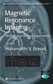
Even as magnetic resonance imaging (MRI) has matured into an invaluable diagnostic tool, it has also become a key experimental tool in biological research, where it demonstrates special sensitivity to a plethora of physiological factors, as well as a capacity for use all the way from cellular suspensions to in vivo human studies. In Magnetic Resonance Imaging: Methods and Biologic Applications, leading experts in the use of MRI from both academia and industry explain its basic principles and demonstrate its power to understand biological processes with numerous cutting-edge applications. To illustrate its capability to reveal exquisite anatomical detail, the authors discuss MRI applications to developmental biology, mouse phenotyping, and fiber architecture. MRI can also provide information about organ and tissue function based on endogenous contrast mechanisms. Examples of brain, kidney, and cardiac function are included, as well as applications to neuro- and tumor pathophysiology. In addition, the volume demonstrates the use of exogenous contrast material in functional assessment of the lung, noninvasive evaluation of tissue pH, the imaging of metabolic activity or gene expression that occur on a molecular level, and cellular labeling using superparamagnetic iron oxide contrast agents. Cutting-edge and user-friendly, Magnetic Resonance Imaging: Methods and Biologic Applications illuminates for biological scientists the basic principles of MRI and shows how it can be used successfully to solve important biological problems.
Radiology, Medical. --- Imaging / Radiology. --- Clinical radiology --- Radiology, Medical --- Radiology (Medicine) --- Medical physics
ISBN: 0521676355 Year: 2006 Publisher: Cambridge : University press,
Abstract | Keywords | Export | Availability | Bookmark
Loading...Choose an application
- Reference Manager
- EndNote
- RefWorks (Direct export to RefWorks)
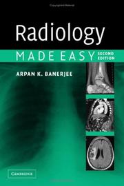
Diagnosis, Differential --- Diagnosis, Differential --- Radiography --- Radiography --- Radiology, medical. --- Methods --- Methods
ISBN: 1280972084 9786610972081 1597458198 1588293572 Year: 2006 Publisher: Totowa, N.J. : Humana Press,
Abstract | Keywords | Export | Availability | Bookmark
Loading...Choose an application
- Reference Manager
- EndNote
- RefWorks (Direct export to RefWorks)
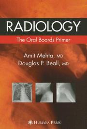
Oral radiology board examinations are notoriously difficult and stressful, requiring a sound working knowledge of all the standard exams and disease pathology encountered in radiology practice. Radiology: The Oral Boards Primer concisely and comprehensively organizes the essential information needed to pass the board exams in diagnostic radiology in an easy to review and easy to memorize format. Drawing on pertinent and key differential diagnoses, the authors have assembled and organized the diagnoses most likely to appear on the exam and illustrated them with essential images to reinforce the findings associated with each differential. Additionally, with each finding set a mnemonic is provided to augment recall of any missing components of the differential that would be considered important. Because of their concise presentation, many cases can be examined, interpreted, completed, and memorized very rapidly in a single sitting. Since the majority of cases contain prototypical representations of pathology, the book also serves as an excellent reference during residency training, as well as a source for many years after the reader has taken and passed the oral board examination. Thoughtfully designed for test success, Radiology: The Oral Boards Primer will greatly assist in the preparation for the American Board of Radiology examination and inspire the assuredness and confidence that comes from being superbly prepared.
Radiology, Medical --- Oral examinations. --- Examinations --- Clinical radiology --- Radiology (Medicine) --- Medical physics --- Radiology, Medical. --- Imaging / Radiology. --- Radiology. --- Radiological physics --- Physics --- Radiation
ISBN: 1281133892 9786611133894 1592599923 1588296709 Year: 2006 Publisher: Totowa, N.J. : Humana Press,
Abstract | Keywords | Export | Availability | Bookmark
Loading...Choose an application
- Reference Manager
- EndNote
- RefWorks (Direct export to RefWorks)
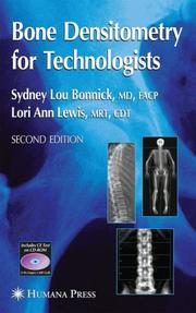
Continuing the excellent reputation of the first edition, leading densitometry experts Sydney Lou Bonnick, MD, FACP, and Lori Ann Lewis, MRT, CDT, have updated and expanded Bone Densitometry for Technologists to reflect the latest standards and developments in the field. Here radiologic technologists, nurse practitioners, physician assistants, and dedicated densitometry technologists can find new guidelines for bone density testing, new therapies for osteoporosis, and new treatment guidelines for osteoporosis, as well as new chapters on pediatric densitometry body composition assessments, and the use of skeletal morphometry in diagnosis and fracture risk prediction. The twelve appendices have also been updated to reflect the most current information available, including contacts for densitometry equipment manufacturers and organizations, guidelines for bone density testing and CPT codes, definitions of terms, and new conversion equations. A companion CD contains the Precision Calculator Companion for automatically calculating the short-term precision and least significant change values for a facility, and the statistical confidence level for any change in BMD, all of which are indispensable in the interpretation of serial bone density studies. In addition, a new continuing education test will allow, upon successful completion, 17 hours of Category A American Society of Radiologic Technologists' credit. Comprehensive and informative, Bone Densitometry for Technologists, Second Edition provides all of the practical knowledge needed to expertly and professionally operate any of today's sophisticated densitometry machines.
Bone densitometry. --- Densitometry, Bone --- Bones --- Calcium in the body --- Radiography --- Radiology, Medical. --- Imaging / Radiology. --- Clinical radiology --- Radiology, Medical --- Radiology (Medicine) --- Medical physics --- Radiology. --- Radiological physics --- Physics --- Radiation
ISBN: 1846283175 1846282969 1846288134 128132891X Year: 2006 Publisher: New York : Springer,
Abstract | Keywords | Export | Availability | Bookmark
Loading...Choose an application
- Reference Manager
- EndNote
- RefWorks (Direct export to RefWorks)
A goldmine of theoretical insights and practical suggestions, Achieving Excellence in Medical Education explores the essential question facing medical educators and learners today: What is our vision of educational excellence, and what can we do to enhance our performance? Among the topics explored within this engaging, informative, and thought-provoking text are: • Education’s position as a priority of medical schools • Seminal educational insights from non-medical educators • Best practices of outstanding educators and learners • Promises and pitfalls of new educational technologies • Key resources for promoting excellence in medical education • Medical education’s role in preparing future leaders • Leadership roles for medical schools in universities and society "Richard Gunderman’s remarkable volume Achieving Excellence in Medical Education is truly a learned treatise on medical education, educational evaluation, academic medical center leadership, and organizational development for excellence." —Thomas S. Inui, ScM, MD, President and CEO, Regenstrief Institute; Sam Regenstrief Professor of Health Services Research; Associate Dean for Health Care Research, and Professor of Medicine, Indiana University School of Medicine, Indianapolis, IN, USA "Gunderman draws on a rich tradition and provocatively challenges us to enact an ethical medicine that makes teaching and learning integral to clinical practice." —Alfred I Tauber, MD, Professor of Philosophy; Zoltan Kohn Professor of Medicine, Director, Center for Philosophy and History of Science, Boston University, Boston Medical Center, Boston, MA, USA.
Medical education. --- Medical personnel --- Professional education --- Education --- Radiology, Medical. --- Medical Education. --- Imaging / Radiology. --- Clinical radiology --- Radiology, Medical --- Radiology (Medicine) --- Medical physics --- Radiology. --- Radiological physics --- Physics --- Radiation
ISBN: 1280804602 9786610804603 0387330488 0387330445 Year: 2006 Publisher: New York : Springer,
Abstract | Keywords | Export | Availability | Bookmark
Loading...Choose an application
- Reference Manager
- EndNote
- RefWorks (Direct export to RefWorks)
The multidetector CT scanner speeds diagnosis and treatment of patients. One of its many uses is to perform CT coronary angiography. Multidetector CT has generated excitement within the cardiology and radiology community as it provides clear pictures and takes less time than other non-invasive techniques, including conventional spiral and electron-beam CT which can take up to an hour or more. This atlas presents over 160 illustrations, with 116 in color and illustrates the capacity of multidetector CT for the analysis of the anatomy of the coronary arteries. Guillem Pons-Llado, MD is the Director of the Cardiac Imaging Unit and at the Hospital de la Santa Creu I Santa Pau, Universitat Autonoma de Barcelona in Barcelona, Spain. Ruben Leta-Petracca, MD is part of the Cardiac Imaging Unit at the Hospital de la Santa Creu I Sant Pau, Universitat Autonoma de Barcelona in Barcelona, Spain.
Heart --- Internal medicine. --- Tomography. --- Medicine, Internal --- Medicine --- Cardiographic tomography --- Diseases --- Tomography --- Radiography --- Cardiology. --- Radiology, Medical. --- Imaging / Radiology. --- Clinical radiology --- Radiology, Medical --- Radiology (Medicine) --- Medical physics --- Internal medicine --- Radiology. --- Radiological physics --- Physics --- Radiation
ISBN: 1280617861 9786610617869 3540282033 3540282025 Year: 2006 Publisher: Berlin ; New York : Springer,
Abstract | Keywords | Export | Availability | Bookmark
Loading...Choose an application
- Reference Manager
- EndNote
- RefWorks (Direct export to RefWorks)
Accelerated partial breast irradiation (APBI) is being rapidly introduced into the clinical management of early breast cancer. APBI, in fact, encompasses a number of different techniques and approaches that include brachytherapy, intraoperative, and external beam techniques. There is currently no single source that describes these techniques and their clinical implementation. This text will be a concise handbook designed to assist the clinician in the implementation of APBI. This will include a review of the principles that underlie APBI, a practical and detailed description of each technique for APBI, a review of current clinical results of APBI, and a review of the incidence and management of treatment related complications.
Breast --- Radiotherapy. --- Cancer --- Radiation therapy --- Electrotherapeutics --- Hospitals --- Medical electronics --- Medical radiology --- Therapeutics, Physiological --- Phototherapy --- Radiological services --- Oncology . --- Radiology, Medical. --- Oncology. --- Diagnostic Radiology. --- Clinical radiology --- Radiology, Medical --- Radiology (Medicine) --- Medical physics --- Tumors --- Radiology. --- Radiological physics --- Physics --- Radiation
Book
ISBN: 1280610298 9786610610297 3540303561 Year: 2006 Publisher: Berlin, Heidelberg : Springer Berlin Heidelberg : Imprint: Springer,
Abstract | Keywords | Export | Availability | Bookmark
Loading...Choose an application
- Reference Manager
- EndNote
- RefWorks (Direct export to RefWorks)
Intensity-modulated radiation therapy (IMRT), one of the most important developments in radiation oncology in the past 25 years, involves technology to deliver radiation to tumors in the right location, quantity and time. Unavoidable irradiation of surrounding normal tissues is distributed so as to preserve their function. The achievements and future directions in the field are grouped in the three sections of the book, each suitable for supporting a teaching course. Part 1 contains topical reviews of the basic principles of IMRT, part 2 describes advanced techniques such as image-guided and biologically based approaches, and part 3 focuses on investigation of IMRT to improve outcome at various cancer sites.
Radiotherapy --- Cancer --- Technological innovations. --- Radiotherapy. --- Radiation therapy --- Electrotherapeutics --- Hospitals --- Medical electronics --- Medical radiology --- Therapeutics, Physiological --- Phototherapy --- Treatment --- Radiological services --- Radiology, Medical. --- Oncology . --- Imaging / Radiology. --- Oncology. --- Tumors --- Clinical radiology --- Radiology, Medical --- Radiology (Medicine) --- Medical physics --- Radiology. --- Radiological physics --- Physics --- Radiation
ISBN: 1280608633 9786610608638 3211322345 321128253X Year: 2006 Publisher: Vienna : Springer-Verlag,
Abstract | Keywords | Export | Availability | Bookmark
Loading...Choose an application
- Reference Manager
- EndNote
- RefWorks (Direct export to RefWorks)
Part of the series sponsored by the European Association of Neurosurgical Societies. The Advances section presents fields of neurosurgery and related areas. The Technical Standards section features detailed descriptions of standard procedures to assist young neurosurgeons in their post-graduate training.
Nervous system --- Surgery. --- Nerves --- Neurosurgery --- Surgery --- Neurosurgery. --- Neurology. --- Radiology, Medical. --- Neurosciences. --- Neuroradiology. --- Neural sciences --- Neurological sciences --- Neuroscience --- Medical sciences --- Clinical radiology --- Radiology, Medical --- Radiology (Medicine) --- Medical physics --- Medicine --- Neuropsychiatry --- Diseases --- Neurology . --- Neuroradiography --- Neuroradiology
ISBN: 3540434674 3540315306 9786610627110 1280627115 3540315551 Year: 2006 Publisher: Berlin, Heidelberg : Springer Berlin Heidelberg : Imprint: Springer,
Abstract | Keywords | Export | Availability | Bookmark
Loading...Choose an application
- Reference Manager
- EndNote
- RefWorks (Direct export to RefWorks)
Magnetic resonance imaging (MRI) has become the leading cross-sectional imaging method in clinical practice. Continuous technical improvements have significantly broadened the scope of applications. At present, MR imaging is not only the most important diagnostic technique in neuroradiology and musculoskeletal radiology, but has also become an invaluable diagnostic tool for abdominal, pelvic, cardiac, breast and vascular imaging. This book offers practical guidelines for performing efficient and cost-effective MRI examinations in daily practice. The underlying idea is that, by adopting a practical protocol-based approach, the work-flow in a MRI unit can be streamlined and optimized. For the second edition, all chapters have been thoroughly reviewed, and new techniques and figures were included. This book will help beginners to advance their starting point in implementing the protocols and will aid more experienced users in updating their knowledge.
Magnetic resonance imaging. --- Radiology, Medical. --- Pediatrics. --- Imaging / Radiology. --- Neuroradiology. --- Paediatrics --- Pediatric medicine --- Medicine --- Children --- Clinical radiology --- Radiology, Medical --- Radiology (Medicine) --- Medical physics --- Diseases --- Health and hygiene --- Radiology. --- Neuroradiography --- Neuroradiology --- Nervous system --- Radiological physics --- Physics --- Radiation
| Listing 1 - 10 of 88 | << page >> |
Sort by
|